

Long-term outcomes of triangle tilt surgery for obstetric brachial plexus injury
- PMID: 20130887
- PMCID: PMC2841265
- DOI: 10.1007/s00383-010-2550-4
Long-term outcomes of triangle tilt surgery for obstetric brachial plexus injury
Abstract
Aim: The purpose of this study was to evaluate long-term shoulder functional outcomes from a triangle tilt procedure on obstetric patients, who initially presented with medial rotation contracture and scapular deformity secondary to obstetric brachial plexus injury.
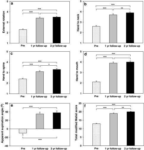
Methods: We retrospectively studied long-term outcomes both functionally and anatomically in 61 patients (age ranging from 2 to 12 years). Functional movements were evaluated and scored using a modified Mallet scale at different time intervals: preoperatively, 1 year and 2 year following triangle tilt surgery. Shoulder anatomy was examined on radiologic images to evaluate the severity of shoulder deformities preoperatively and anatomical improvement after the surgery.
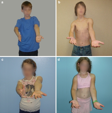
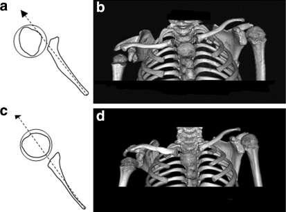
Results: All shoulder functional movements were significantly improved at 1 and 2 year follow-ups. Functional improvements were maintained in shoulder abduction, external rotation and hand-to-mouth movements beyond the first year, and continued in hand-to-neck and hand-to-spine movements past 2 years. Remarkable glenohumeral remodeling or reservation of glenoid congruence was observed in all patients over a mean time of 27 months postoperatively.
Conclusion: The triangle tilt procedure, which addresses scapular and glenohumeral joint abnormalities characteristic of Erb's palsy, improves shoulder functional movements and anatomical structure in patients over the long-term.
Figures

Similar articles
-
What Range of Motion is Achieved 5 Years After External Rotationplasty of the Shoulder in Infants with an Obstetric Brachial Plexus Injury?Clin Orthop Relat Res. 2020 Jan;478(1):114-123. doi: 10.1097/CORR.0000000000000996. Clin Orthop Relat Res. 2020. PMID: 31651590 Free PMC article.
-
The early effects of tendon transfers and open capsulorrhaphy on glenohumeral deformity in brachial plexus birth palsy. Surgical technique.J Bone Joint Surg Am. 2009 Oct 1;91 Suppl 2:213-22. doi: 10.2106/JBJS.I.00501. J Bone Joint Surg Am. 2009. PMID: 19805585
-
Glenohumeral abduction contracture in children with unresolved neonatal brachial plexus palsy.J Bone Joint Surg Am. 2015 Jan 21;97(2):112-8. doi: 10.2106/JBJS.N.00203. J Bone Joint Surg Am. 2015. PMID: 25609437
-
Shoulder problems in children with brachial plexus birth palsy: evaluation and management.J Am Acad Orthop Surg. 2009 Apr;17(4):242-54. doi: 10.5435/00124635-200904000-00005. J Am Acad Orthop Surg. 2009. PMID: 19307673 Review.
-
Arthroscopic treatment for internal contracture of the shoulder secondary to brachial plexus birth palsy: report of a case series and review of the literature.Eur J Orthop Surg Traumatol. 2015 Oct;25(7):1121-9. doi: 10.1007/s00590-015-1670-x. Epub 2015 Jul 14. Eur J Orthop Surg Traumatol. 2015. PMID: 26169993 Review.
Cited by
-
Triangle tilt surgery as salvage procedure for failed shoulder surgery in obstetric brachial plexus injury.Pediatr Surg Int. 2010 Sep;26(9):913-8. doi: 10.1007/s00383-010-2673-7. Epub 2010 Jul 29. Pediatr Surg Int. 2010. PMID: 20668864 Free PMC article.
-
Outcomes of Brachial Plexus Neurolysis in 40 Patients With Obstetric Brachial Plexus Injury.Eplasty. 2025 Jan 29;25:e4. eCollection 2025. Eplasty. 2025. PMID: 40469489 Free PMC article.
-
Triangle tilt and steel osteotomy: similar approaches to common problems.Open Orthop J. 2011 Mar 24;5:124-33. doi: 10.2174/1874325001105010124. Open Orthop J. 2011. PMID: 21584207 Free PMC article.
-
Extended long-term (5 years) outcomes of triangle tilt surgery in obstetric brachial plexus injury.Open Orthop J. 2013 Apr 29;7:94-8. doi: 10.2174/1874325001307010094. Print 2013. Open Orthop J. 2013. PMID: 23730369 Free PMC article.
-
10-year Follow-up of Mod Quad and Triangle Tilt Surgeries in Obstetric Brachial Plexus Injury.Plast Reconstr Surg Glob Open. 2019 Jan 22;7(1):e1998. doi: 10.1097/GOX.0000000000001998. eCollection 2019 Jan. Plast Reconstr Surg Glob Open. 2019. PMID: 30859023 Free PMC article.
References
-
- Birch R (2001) Late sequelae at the shoulder in obstetrical palsy in children. In: Randelli M, Karlsson J (eds) Surgical techniques in orthopaedics and traumatology: shoulder, Elsevier, Paris, pp 55-200-E-210
MeSH terms
LinkOut - more resources
Full Text Sources
Medical
Miscellaneous
