Review
. 2010 Jun;25(6):1017-23.
doi: 10.1007/s00467-009-1425-1. Epub 2010 Feb 4.

Enzymatic disease of the podocyte

Affiliations
Review

Enzymatic disease of the podocyte

Andreas D Kistler et al. Pediatr Nephrol. 2010 Jun.

Abstract

Proteinuria is an early sign of kidney disease and has gained increasing attention over the past decade because of its close association with cardio-vascular and renal morbidity and mortality. Podocytes have emerged as the cell type that is critical in maintaining proper functioning of the kidney filter. A few genes have been identified that explain genetic glomerular failure and recent insights shed light on the pathogenesis of acquired proteinuric diseases. This review highlights the unique role of the cysteine protease cathepsin L as a regulatory rather than a digestive protease and its action on podocyte structure and function. We provide arguments why many glomerular diseases can be regarded as podocyte enzymatic disorders.


Figures

Fig. 1
Fig. 1
Schematic of CatL mRNA containing several AUG codons and resulting proteins. After translation from the first AUG, CatL is processed to yield a 30-kDa lysosomal form, called single-chain CatL (black arrows). However, alternative translation initiation from a downstream AUG produces a CatL isoform devoid of the lysosomal targeting sequence (short CatL), which localizes to the cytoplasm (red arrow). Adapted with permission from Sever et al. [15]
Fig. 2
Fig. 2
Cathepsin L (CatL) is essential for the development of proteinuria in the lipopolysaccharide (LPS) model. In wild-type (WT) mice, intraperitoneal injection of LPS leads to a T- and B-cell-independent transient form of proteinuria through the activation of podocyte TLR-4 and induction of B7-1 [39]. a Immunocytochemistry of mouse glomeruli using monoclonal anti-CatL antibody. WT mice receiving LPS (WT LPS) upregulate the expression of cytosolic CatL compared with control mice receiving PBS (WT CON). LPS was also injected into CatL−/− mice (CatL−/− LPS). Original magnification, ×400. b Electron micrographs of foot processes (FPs) showing effacement in LPS-treated WT, but not in CatL−/− mice. Adapted with permission from Sever et al. [15]
Fig. 3
Fig. 3
Cathepsin L (CatL) mRNA and protein expression are elevated in human proteinuric kidney diseases. a Quantitative rt-PCR of microdissected glomeruli from human biopsies of patients with acquired proteinuric diseases: minimal change disease (MCD; n=7), membranous glomerulonephritis (MGN; n=9), focal segmental glomerulosclerosis (FSGS; n=7), and diabetic nephropathy (DN; n=10). **P<0.01 for comparison with healthy controls (CON; n=8). b CatL labeling of normal human kidney. c CatL labeling of a kidney biopsy from a patient with diabetic nephropathy, mildly reduced renal function, and nephrotic range proteinuria. Adapted with permission from Sever et al. [15]
Fig. 4
Fig. 4
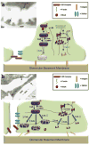
Cathepsin L (CatL) is induced in podocytes during FP effacement. a Schematic illustration of FP effacement and proteinuria as a podocyte enzymatic disease. Under normal conditions, synaptopodin and dynamin are involved in regulating podocyte F-actin. A small portion of CatL is in the cytosol and participates in a physiological turnover of synaptopodin and dynamin. The electron micrograph of control mice (insert), shows CatL expression located mainly in lysosomes of primary podocyte processes (dashed arrow). Only few gold labeling is found in FP (solid arrows). b The induction of cytosolic CatL causes proteolysis of synaptopodin and dynamin, thereby disrupting actin organization, causing podocyte FP effacement and proteinuria. The electron micrograph of LPS treated mice (insert) shows FP effacement and induction of CatL in lysosomes of primary processes (dashed arrow) and in effaced podocyte FPs (solid arrows); CaN calcineurin; PKA protein kinase A; CaMKII calcium-dependent protein kinase II; P podocyte; GBM glomerular basement membrane; END endothelial cells; ERY erythrocyte

References

    1. Hogg RJ, Portman RJ, Milliner D, Lemley KV, Eddy A, Ingelfinger J. Evaluation and management of proteinuria and nephrotic syndrome in children: recommendations from a pediatric nephrology panel established at the National Kidney Foundation conference on proteinuria, albuminuria, risk, assessment, detection, and elimination (PARADE) Pediatrics. 2000;105:1242–1249. - PubMed
    1. Pavenstadt H, Kriz W, Kretzler M. Cell biology of the glomerular podocyte. Physiol Rev. 2003;83:253–307. - PubMed
    1. Reiser J, Kriz W, Kretzler M, Mundel P. The glomerular slit diaphragm is a modified adherens junction. J Am Soc Nephrol. 2000;11:1–8. - PubMed
    1. Patrakka J, Tryggvason K. New insights into the role of podocytes in proteinuria. Nat Rev Nephrol. 2009;5:463–468. - PubMed
    1. Reiser J, Oh J, Shirato I, Asanuma K, Hug A, Mundel TM, Honey K, Ishidoh K, Kominami E, Kreidberg JA, Tomino Y, Mundel P. Podocyte migration during nephrotic syndrome requires a coordinated interplay between cathepsin L and alpha3 integrin. J Biol Chem. 2004;279:34827–34832. - PubMed

Publication types