Review
. 2010 Jun;104(2-3):245-55.
doi: 10.1007/s11120-010-9533-0. Epub 2010 Feb 4.

The chlorosome: a prototype for efficient light harvesting in photosynthesis

Affiliations
Review

The chlorosome: a prototype for efficient light harvesting in photosynthesis

Gert T Oostergetel et al. Photosynth Res. 2010 Jun.

Abstract

Three phyla of bacteria include phototrophs that contain unique antenna systems, chlorosomes, as the principal light-harvesting apparatus. Chlorosomes are the largest known supramolecular antenna systems and contain hundreds of thousands of BChl c/d/e molecules enclosed by a single membrane leaflet and a baseplate. The BChl pigments are organized via self-assembly and do not require proteins to provide a scaffold for efficient light harvesting. Their excitation energy flows via a small protein, CsmA embedded in the baseplate to the photosynthetic reaction centres. Chlorosomes allow for photosynthesis at very low light intensities by ultra-rapid transfer of excitations to reaction centres and enable organisms with chlorosomes to live at extraordinarily low light intensities under which no other phototrophic organisms can grow. This article reviews several aspects of chlorosomes: the supramolecular and molecular organizations and the light-harvesting and spectroscopic properties. In addition, it provides some novel information about the organization of the baseplate.


Figures

Fig. 1
Fig. 1
Examples of isolated chlorosomes differing in overall shape and size. Specimens were prepared by negative stain embedding with uranyl acetate. a Ellipsoid-shaped chlorosomes of Chlorobaculum tepidum wild-type, the model organism of the green sulphur bacteria. b Conically shaped chlorosomes of Chlorobaculum tepidum bchQRU mutant. c Irregularly shaped chlorosomes with a somewhat undulating surface of Cab. thermophilum, a newly discovered phototrophic microorganism belonging to the Acidobacteria. Size bar for all frames equals 100 nm
Fig. 2
Fig. 2
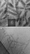
Isolated chlorosomes embedded in an amorphous ice layer give hints of the overall and internal structure. a Overview of unstained chlorosomes of Chlorobium tepidum. The inset shows a fine parallel spacing of lamellae, its calculated diffraction pattern indicates a strong diffraction spot equivalent with a 2.1-nm lamellar spacing. b Unstained ice-embedded chlorosomes of Chloroflexus aurantiacus (phylum Chloroflexi or filamentous anoxygenic phototrophs). The ice layer has been prepared over a holey-carbon film, which is visible at the lower left side. Size bar for both frames equals 100 nm
Fig. 3
Fig. 3
End-on views of chlorosomes of Chlorobaculum tepidum, fixed in a vertical position in an amorphous ice layer. Cryo-EM reveals the packing of the lamellae. a Packing in the wild-type with some of the lamellae in concentric rings, others in a more irregular association. b Packing in the bchQRU mutant, showing a more regular multi-cylindrical organization. See also (Oostergetel et al. 2007) for further images. Size bar equals 25 nm
Fig. 4
Fig. 4
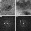
Analysis of the interior of the chlorosome of Chlorobaculum tepidum. a Image of an unstained, ice-embedded chlorosome from the wild-type. b Calculated diffraction pattern from the image of frame a. A bright but unsharp reflection spot (white arrow) indicates an average spacing between lamellae of 2.1 nm, which is also directly visible in the image of frame a. A sharp layer line at 1.25 nm (red arrow) indicates a specific internal repeating distance of 1.25 nm of the lamellae, caused by a specific packing of BChls. A thin but distinct reflection at 3.3 nm (green arrow) is assigned to a spacing of protein molecules of the baseplate. c Image of an unstained, ice-embedded chlorosome from the bclQRU mutant. d Calculated diffraction pattern from the image of c. The white and green arrows indicate structural elements as in the pattern of frame b. The sharp layer line (red arrow) now indicates a specific internal repeating distance of 0.83 nm, instead of 1.25 nm as in the wild-type. The yellow arrow marks a sharp reflection that hints at another type of spacing (1.1 nm), likely in the baseplate. Size bar equals 50 nm for frames a and c
Fig. 5
Fig. 5
Cryo-EM of Chlorobaculum tepidum chlorosomes. a A wild-type chlorosome recorded in an about vertical position (side view), and in a specific angular orientation in which rows of proteins of the baseplate become visible. b Diffraction pattern of a selected part of the chlorosome of frame a, showing that the elements at the edge have a repeating distance of 3.3 nm (white arrows). c A wild-type chlorosome in about horizontal position (top view). The baseplate element is not directly visible because of strong overlap with the interior. d Diffraction pattern of the chlorosome of frame c, showing the same distance of 3.3 nm of elements as in frame b. G.T. Oostergetel, unpublished data). Size bar equals 50 nm
Fig. 6
Fig. 6
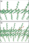
Molecular models of BChl syn-anti monomer stacks in tubular models of a a single stack showing the farnesyl tails alternately extending on both sides. Radius of curvature 10.2 nm. b Two syn-anti stacks interconnected by hydrogen bonds (black dotted line in the centre). The orange arrow indicates the direction of the exciton delocalization pathway over neighbouring stacks along the connecting hydrogen bonds. The models were made in Swiss-PDB Viewer and visualized using Pymol
Fig. 7
Fig. 7
Cylindrical model of the packing of concentric lamellae in the Chlorobaculum tepidum bchQRU mutant, based on distances as observed by electron microscopy and solid-state NMR spectroscopy (Ganapathy et al. 2009). The spacing between layers is 2.1 nm. The green band indicates the position of individual Bchl molecules in four stacks of syn-anti dimers. In the wild-type chlorosomes, the stacks run in the direction of the cylinder axis

Similar articles

Cited by

References

    1. Arellano JB, Melo TB, Borrego CM, Garcia-Gil J, Naqvi KR. Nanosecond laser photolysis studies of chlorosomes and artificial aggregates containing bacteriochlorophyll e: Evidence for the proximity of carotenoids and bacteriochlorophyll a in chlorosomes from Chlorobium phaeobacteroides strain CL1401. Photochem Photobiol. 2000;72:669–675. doi: 10.1562/0031-8655(2000)072<0669:NLPSOC>2.0.CO;2. - DOI - PubMed
    1. Arellano JB, Melo TB, Borrego CM, Naqvi KR. Bacteriochlorophyll e monomers, but not aggregates, sensitize singlet oxygen: implications for a self-photoprotection mechanism in chlorosomes. Photochem Photobiol. 2002;76:373–380. doi: 10.1562/0031-8655(2002)076<0373:BEMBNA>2.0.CO;2. - DOI - PubMed
    1. Balaban TS, Tamiaki H, Holzwarth AR. Chlorins programmed for self-assembly. Top Curr Chem. 2005;258:1–38. doi: 10.1007/b137480. - DOI
    1. Blankenship RE, Matsuura K (2003) Antenna complexes from green photosynthetic bacteria. In: Green BR, Parson WW (eds) Light-harvesting antennas in photosynthesis. Kluwer Academic Publishers, Dordrecht, pp 195–217
    1. Blankenship RE, Olson JM, Miller M (1995) Antenna complexes from green photosynthetic bacteria. In: Blankenship RE, Madigan MT, Bauer CE (eds) Anoxygenic photosynthetic bacteria. Kluwer Academic Publishers, Dordrecht, pp 399–435

Publication types

Substances

LinkOut - more resources