. 2010 Mar;120(3):546-51.
doi: 10.1002/lary.20797.

Characterization of mesenchymal stem cells from human vocal fold fibroblasts

Affiliations

Characterization of mesenchymal stem cells from human vocal fold fibroblasts

Summer E Hanson et al. Laryngoscope. 2010 Mar.

Abstract

Objectives/hypothesis: Mesenchymal stem cells (MSCs) originally isolated from bone marrow (BM), are fibroblast-looking cells that are now assumed to be present in the stromal component of many tissues. MSCs are characterized by a certain set of criteria, including their growth culture characteristics, a combination of cell surface markers, and the ability to differentiate along multiple mesenchymal tissue lineages. We hypothesized that human vocal fold fibroblasts (hVFF) isolated from the lamina propria meet the criteria established to define MSCs and are functionally similar to MSCs derived from BM and adipose tissue.

Study design: In vitro study.

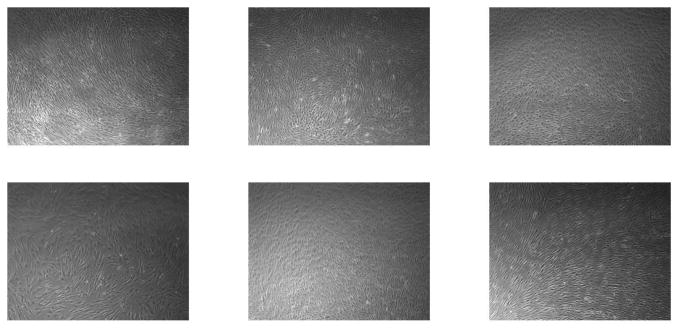
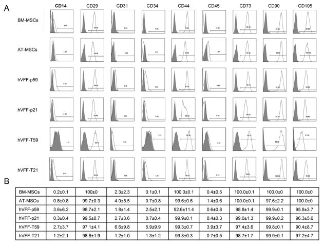
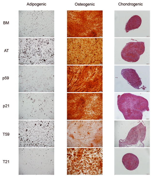
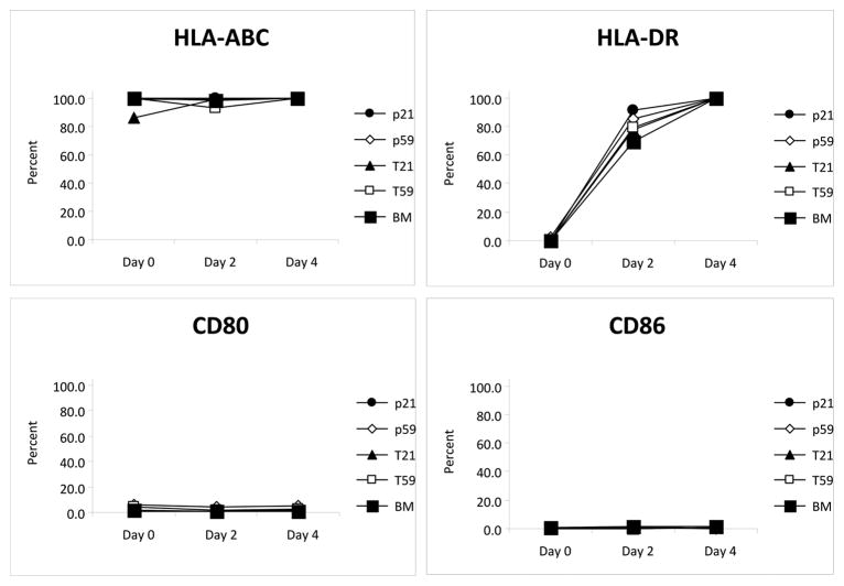
Methods: hVFF were previously derived from human vocal fold tissues. MSCs were derived from adipose tissue (AT), and BM of healthy donors based on their attachment to culture dishes and their morphology and expanded in culture. Cells were analyzed for standard cell surface markers identified on BM-derived MSCs and the ability to differentiate into cells of mesenchymal lineage (i.e., fat, bone, and cartilage). We investigated the immunophenotype of these cells before and after interferon-gamma (INF-gamma) stimulation.

Results: hVFF displayed cell surface markers and multipotent differentiation capacity characteristic of MSCs. Furthermore, these cells exhibited similar patterns of expression of human leukocyte antigen and costimulatory molecules, after stimulation with INF-gamma compared to MSCs derived from BM and AT.

Conclusions: Based on our findings, hVFF derived from lamina propria have the same cell surface markers, immunophenotypic characteristics, and differentiation potential as BM- and AT-derived MSCs. We propose that vocal fold fibroblasts are MSCs resident in the vocal fold lamina propria.


Conflict of interest statement

Conflict of Interest: None

Figures

Figure 1
Figure 1. Morphology of hVFF
Representative microscopic views (5X) of human bone marrow-derived mesenchymal stem cells (MSCs) (upper left), human adipose tissue-derived MSCs (lower left); human vocal fold fibroblasts (hVFF) p59 (upper middle); hVFF p21 (lower middle); hVFF T59 (upper right); hVFF T21 (lower right).
Figure 2
Figure 2. Phenotype of hVFF
Panel A shows representative fluorescent activated cell sorting analysis of hVFF cell lines p59, p21, T59 and T21 compared to BM- and AT- derived MSCs for different cell surface markers. Panel B shows a table of the mean value (± standard error) of the expression of corresponding markers. These experiments were performed in triplicate.
Figure 3
Figure 3. Differentiation Potential
Representative microscopic images views of the differentiation of hVFF cell lines p59, p21, T59 and T21 compared to BM- and AT derived MSCs into adipogenic (left column), osteogenic (middle column), and chondrogenic lineages (right column). Oil red O staining shows lipid vacuoles stained red; Alizarin red S staining shows deposits of calcium crystals stained orange to brown, and Safranin O staining shows cartilage-specific glycosaminoglycans.
Figure 4
Figure 4. Immunophenotype of hVFF
Unstimulated BM-MSCs and hVFF cells are positive for HLA-ABC but do not express HLA-DR, CD80 or CD86. Treatment of cells with IFN-γ induces the cell surface expression of HLA-DR while CD80 and CD86 remain unchanged. Panel A shows the trend in cell surface marker expression over four days of treatment with IFN-γ for BM-MSCs, hVFF-p59, hVFF-p21, hVFF-T59 and hVFF-T21. Panel B shows a table of the mean value (± standard error) of the expression of surface markers. These experiments were performed in triplicate.

Similar articles

Cited by

References

    1. Hematti P. Role of mesenchymal stromal cells in solid organ transplantation. Transplant Rev (Orlando) 2008;22(4):262–273. - PMC - PubMed
    1. Trivedi P, Hematti P. Derivation and immunological characterization of mesenchymal stromal cells from human embryonic stem cells. Exp Hematol. 2008;36(3):350–359. - PMC - PubMed
    1. Thibeault SL, Klemuk SA, Smith ME, Leugers C, Prestwich G. In Vivo Comparison of Biomimetic Approaches for Tissue Regeneration of the Scarred Vocal Fold. Tissue Eng Part A. 2008 - PMC - PubMed
    1. Thibeault SL, Li W, Bartley S. A method for identification of vocal fold lamina propria fibroblasts in culture. Otolaryngol Head Neck Surg. 2008;139(6):816–822. - PMC - PubMed
    1. Hansen JK, Thibeault SL, Walsh JF, Shu XZ, Prestwich GD. In vivo engineering of the vocal fold extracellular matrix with injectable hyaluronic acid hydrogels: early effects on tissue repair and biomechanics in a rabbit model. Ann Otol Rhinol Laryngol. 2005;114(9):662–670. - PubMed

Publication types

Substances

LinkOut - more resources