. 2010 May;51(5):1778-88.
doi: 10.1002/hep.23511.

Mammalian target of rapamycin regulates vascular endothelial growth factor-dependent liver cyst growth in polycystin-2-defective mice

Affiliations

Mammalian target of rapamycin regulates vascular endothelial growth factor-dependent liver cyst growth in polycystin-2-defective mice

Carlo Spirli et al. Hepatology. 2010 May.

Abstract

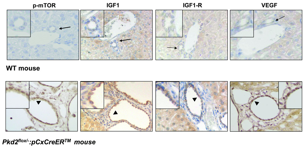
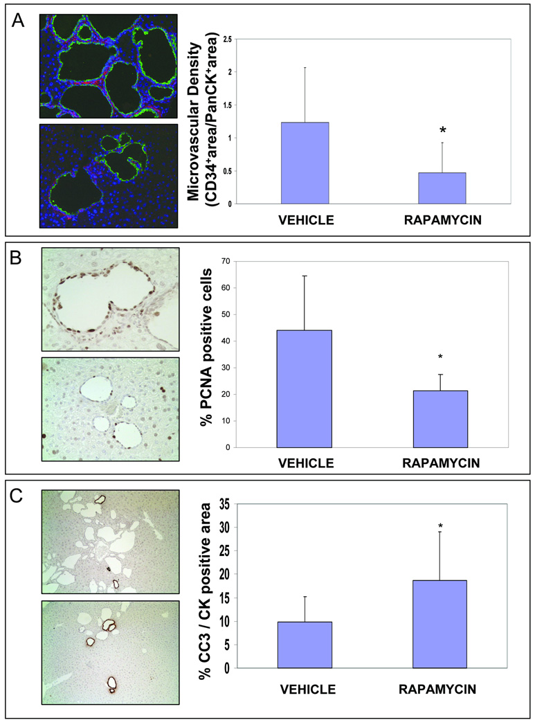
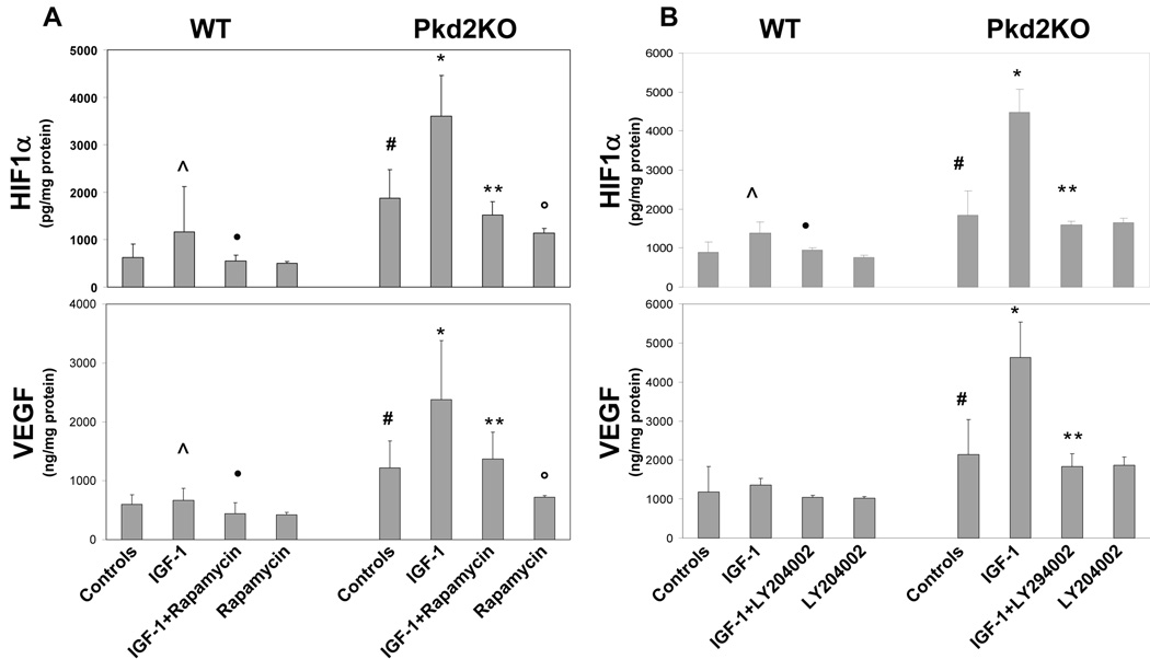
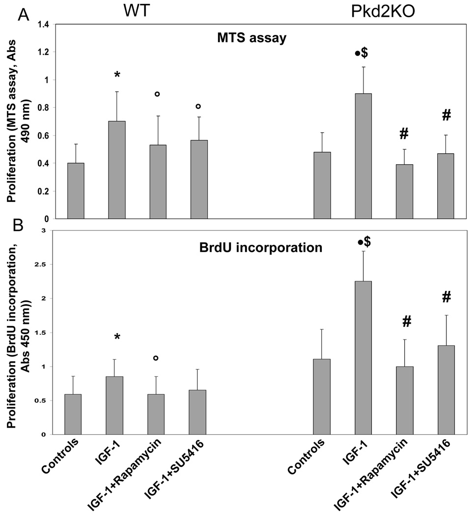
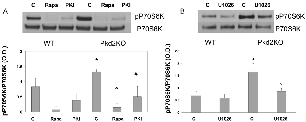
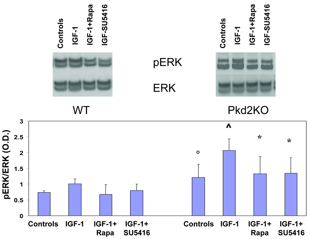
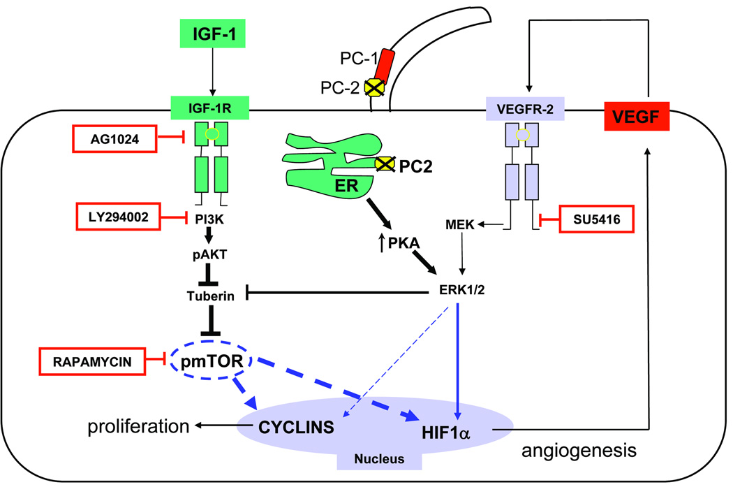
Polycystic liver disease may complicate autosomal dominant polycystic kidney disease (ADPKD), a disease caused by mutations in polycystins, which are proteins that regulate signaling, morphogenesis, and differentiation in epithelial cells. The cystic biliary epithelium [liver cystic epithelium (LCE)] secretes vascular endothelial growth factor (VEGF), which promotes liver cyst growth via autocrine and paracrine mechanisms. The expression of insulin-like growth factor 1 (IGF1), insulin-like growth factor 1 receptor (IGF1R), and phosphorylated mammalian target of rapamycin (p-mTOR) and the protein kinase A (PKA)-dependent phosphorylation of extracellular signal-regulated kinase 1/2 (ERK1/2) are also up-regulated in LCE. We have hypothesized that mammalian target of rapamycin (mTOR) represents a common pathway for the regulation of hypoxia-inducible factor 1 alpha (HIF1alpha)-dependent VEGF secretion by IGF1 and ERK1/2. Conditional polycystin-2-knockout (Pkd2KO) mice were used for in vivo studies and to isolate cystic cholangiocytes [liver cystic epithelial cells (LCECs)]. The expression of p-mTOR, VEGF, cleaved caspase 3 (CC3), proliferating cell nuclear antigen (PCNA), IGF1, IGF1R, phosphorylated extracellular signal-regulated kinase, p-P70S6K, HIF1alpha, and VEGF in LCE, LCECs, and wild-type cholangiocytes was studied with immunohistochemistry, western blotting, or enzyme-linked immunosorbent assays. The cystic area was measured by computer-assisted morphometry of pancytokeratin-stained sections. Cell proliferation in vitro was studied with 3-(4,5-dimethylthiazol-2-yl)-5-(3-carboxymethoxyphenyl)-2-(4-sulfophenyl)-2H-tetrazolium and bromodeoxyuridine assays. The treatment of Pkd2KO mice with the mTOR inhibitor rapamycin significantly reduced the liver cyst area, liver/body weight ratio, pericystic microvascular density, and PCNA expression while increasing expression of CC3. Rapamycin inhibited IGF1-stimulated HIF1alpha accumulation and VEGF secretion in LCECs. IGF1-stimulated LCEC proliferation was inhibited by rapamycin and SU5416 (a vascular endothelial growth factor receptor 2 inhibitor). Phosphorylation of the mTOR-dependent kinase P70S6K was significantly reduced by PKA inhibitor 14-22 amide and by the mitogen signal-regulated kinase inhibitor U1026.

Conclusion: These data demonstrate that PKA-dependent up-regulation of mTOR has a central role in the proliferative, antiapoptotic, and pro-angiogenic effects of IGF1 and VEGF in polycystin-2-defective mice. This study also highlights a mechanistic link between PKA, ERK, mTOR, and HIF1alpha-mediated VEGF secretion and provides a proof of concept for the potential use of mTOR inhibitors in ADPKD and conditions with aberrant cholangiocyte proliferation.


Conflict of interest statement

All Authors state there are no conflicts of interest to disclose

Figures

Figure 1
Figure 1. Expression of p-mTOR, IGF1, IGF1-R and VEGF in cystic cholangiocytes of Pkd2KO mice
Paraffin embedded liver sections (5µM) of WT and Pkd2flox/−:pCxCreERTM (Pkd2KO) mice were labeled with specific antibodies against the phosphorylated form of mTOR, IGF1, IGF1-R and VEGF. Immunoreactivity for p-mTOR IGF1, IGF1-R and VEGF (arrowhead) is up-regulated on the biliary epithelium in Pkd2KO mice, as respect to control mice (arrow). Magnification: 40×
Figure 2
Figure 2. Rapamycin reduces cystic area and liver weight/BW percentage in Pkd2KO mice
(A) Micrographs are representative of vehicle (left) and rapamycin (1.5 mg/Kg/day) treated mice (right). (B) As shown in the bar graph, a significant reduction in cystic area was observed in Pkd2KO treated animals (°=p<0.001, n=10). (C) The decrease in cysts growth is reflected also in the significant reduction of liver weight/body weight ratio. In fact, liver weight/body weight ratio was higher in mice treated with vehicle (n=10) compared to wild type mice (**p<0.001, n=6) and was significantly reduced in rapamycin treated mice (n=10) (*=p<0.01).
Figure 3
Figure 3. Rapamycin reduces microvascular density (CD34) and cell proliferation (PCNA), while increases apoptosis (CC3 expression) and in Pkd2KO mice
Each couple of micrographs is representative of vehicle (above) and rapamycin treated mice (below). For quantitative analysis five random non-overlapping fields taken at 40× magnification per each slide were recorded by a digital camera connected to Spot Advanced imaging software (version 3.5) by an observer who was blinded to the treatment modality. (A) Rapamycin treatment reduced microvascular density in Pkd2KO mice. Paraffin embedded liver sections (5µM) were stained with an anti-CD34 and with an anti-Cow Cytokeratin Wide Spectrum (PanCK). To calculate the vascular and biliary areas, two different thresholds were set out for CD34 (red fluorescence) and PanCK (green fluorescence) positive structures respectively, and then expressed as percentage of pixel above the threshold per field. As shown in the bar graphs treatment with rapamycin significantly reduced the microvascular density. (# = p<0.05 vs vehicle;). (B) Cystic cholangiocytes showed strong proliferative activity (PCNA staining). As shown in the bar graph, a significant reduction in PCNA expression, assessed by morphometric analysis, was observed in Pkd2KO treated animals (*=p<0.001 n=10). An average of 1000 nuclei was counted per each mouse and the percentage of PCNA positive nuclei was then calculated. Only strongly positive immunostained nuclei were considered as PCNA positive. (B) To account for the decrease in liver cysts in rapamycin-treated mice, we quantified the amount of apoptosis (Cleaved Caspase-3 staining) in cystic cells. The bar graph show a significant increase in CC3 expression in Pkd2KO treated animals (*=p<0.01, n=10). CC3-positive area was expressed as percent of the cytokeratin-positive area.
Figure 4
Figure 4. Rapamycin and LY294002 inhibited IGF1-induced HIF1α accumulation and VEGF secretion
The effect of rapamycin on VEGF secretion and on the nuclear expression of its main transcription factor, HIF1α, in primary cultures of cystic cholangiocytes cultured from Pkd2KO mice was assessed by ELISA. (A) In Pkd2KO cultured cholangiocytes, HIF-1α accumulation and VEGF secretion induced by IGF1 are significantly higher as respect to WT cholangiocytes. This effect was completely blunted in cells treated with rapamycin (5 µM) (n=4) and in cells treated with a PI3K inhibitor, LY204002 (10 µM) (n=3) (B). (#= p<0.005 vs WT C), (^= p<0.05 vs WT C), (•= p<0.05 vs WT+IGF-1), (*= p<0.001 vs Pkd2KO C), (**= p<0.001 vs Pkd2KO+IGF-1)
Figure 5
Figure 5. Rapamycin and SU5614 inhibit IGF-1-induced cell proliferation in cultured Pkd2KO cystic cholangiocytes
Using two different cell proliferation assays similar results were obtained. MTS assay results are shown in panel A, and BrdU incorporation is shown in panel B. In both WT and Pdk2KO cholangiocytes, IGF1 significantly enhanced cell proliferation compared to untreated cells (*=p<0.05 with respect to WT controls cholangiocytes. •=p<0.01 with respect to control Pkd2KO cholangiocytes.) The increase in cell proliferation stimulated by Pkd2KO cholangiocytes was significantly higher in Pkd2KO cholangiocytes (&=p<0.05) with respect to WT cholangiocytes exposed to IGF-1). IGF1-induced cell proliferation was significantly inhibited by treatment with rapamycin (5 µM) or with SU5416, a VEGFR-2 inhibitor; °=p<0.05 in WT cholangiocytes treated with IGF-1 plus rapamycin or SU5416, as respect to IGF-1-treated WT cholangiocytes; #=p<0.01 in Pkd2KO cholangiocytes treated with IGF-1 plus rapamycin or SU5416, as respect to IGF-1-treated Pkd2KO cholangiocytes;) (n=3).
Figure 6
Figure 6. Phosphorylation of the mTOR-activated P70S6K is PKA- and ERK-mediated
A) In Pkd2KO cystic cholangiocytes the ratio in phospho-P70S6K/P70S6K is higher with respect to WT cells, it was completely inhibited by rapamycin, as expected, and significantly inhibited by the PKA inhibitor PKI (*=p<0.05 with respect to WT untreated cholangiocytes; ^=p<0.001 with respect to Pkd2KO untreated cholangiocytes; #=p<0.01 with respect to Pkd2KO untreated cholangiocytes). B) In Pkd2KO cholangiocytes the ratio in phospho-P70S6K/P70S6K was significantly inhibited by the MEK inhibitor U1026 (10µM). (*=p<0.05 with respect to WT untreated cholangiocytes; °=p<0.05 with respect to Pkd2KO untreated cholangiocytes)
Figure 7
Figure 7. Rapamycin and SU5614 inhibit IGF-1-induced ERK phosphorylation in cultured Pkd2KO cystic cholangiocytes
IGF1 significantly enhances ERK phosphorylation. IGF1-induced ERK phosphorylation was significantly inhibited by treatment with rapamycin (5 µM) or with SU5416, a VEGFR-2 inhibitor; the bar graph illustrates the ratio in phosphorylated ERK/ERK as assessed by densitometry (°=p<0.05 as respect to WT cells) (^=p<0.05 as respect to Pkd2KO controls) (*p<0.05 as respect to Pkd2KO controls) (n=4).
Figure 8
Figure 8. Working Model to explain cyst growth in PC2-defective cholangiocytes
The cartoon summarizes the main findings of this study, in the contest of the most recent literature, . Cells with defective PC2 are characterized by increased PKA production and ERK1/2 phosphorylation. Erk 1/2 stimulates HIF1α-dependent VEGF secretion directly and by inhibiting tuberin, a negative regulator of mTOR. mTOR has a central role in IGF-1-stimulated proliferation of cystic cholangiocytes. IGF-1, a growth factor secreted by the cystic epithelium and by cholangiocyte under stress, binds to its receptor IGF1-R and activates the PI3K/AKT/mTOR pathway; mTOR stimulates proliferation through cyclins and through a HIF1α/VEGF-dependent autocrine loop. Rapamycin is an mTOR inhibitor; LY294002 is a PI3K inhibitor; AG1024 is an IGF-1R inhibitor; SU5416 is a VEGFR-2 inhibitor

Similar articles

Cited by

References

    1. Bae KT, Zhu F, Chapman AB, Torres VE, Grantham JJ, Guay-Woodford LM, et al. Magnetic resonance imaging evaluation of hepatic cysts in early autosomal-dominant polycystic kidney disease: the Consortium for Radiologic Imaging Studies of Polycystic Kidney Disease cohort. Clin J Am Soc Nephrol. 2006;1:64–69. Epub 2005 Oct 26. - PubMed
    1. Torres VE, Harris PC. Mechanisms of Disease: autosomal dominant and recessive polycystic kidney diseases. Nat Clin Pract Nephrol. 2006;2:40–55. quiz 55. - PubMed
    1. Anyatonwu GI, Estrada M, Tian X, Somlo S, Ehrlich BE. Regulation of ryanodine receptor-dependent calcium signaling by polycystin-2. Proc Natl Acad Sci U S A. 2007;104:6454–6459. Epub 2007 Apr 2. - PMC - PubMed
    1. Li Y, Wright JM, Qian F, Germino GG, Guggino WB. Polycystin 2 interacts with type I inositol 1,4,5-trisphosphate receptor to modulate intracellular Ca2+ signaling. J Biol Chem. 2005;280:41298–41306. Epub 2005 Oct 13. - PubMed
    1. Alvaro D, Onori P, Alpini G, Franchitto A, Jefferson DM, Torrice A, et al. Morphological and functional features of hepatic cyst epithelium in autosomal dominant polycystic kidney disease. Am J Pathol. 2008;172:321–332. - PMC - PubMed

Publication types

MeSH terms

Substances