. 2010 Mar 17;21(3):436-44.
doi: 10.1021/bc9003102. Epub 2010 Feb 4.

A dual-labeled knottin peptide for PET and near-infrared fluorescence imaging of integrin expression in living subjects

Affiliations

A dual-labeled knottin peptide for PET and near-infrared fluorescence imaging of integrin expression in living subjects

Richard H Kimura et al. Bioconjug Chem. .

Abstract

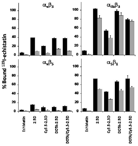
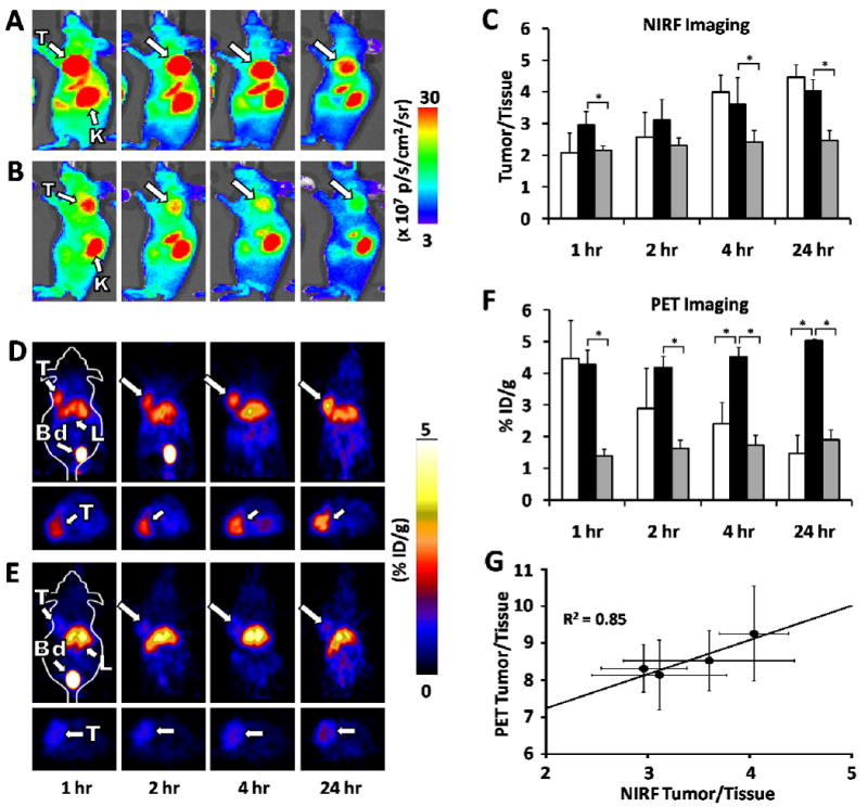
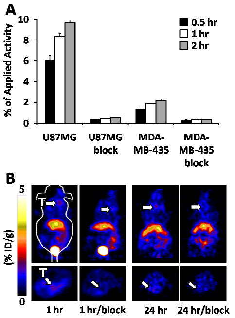
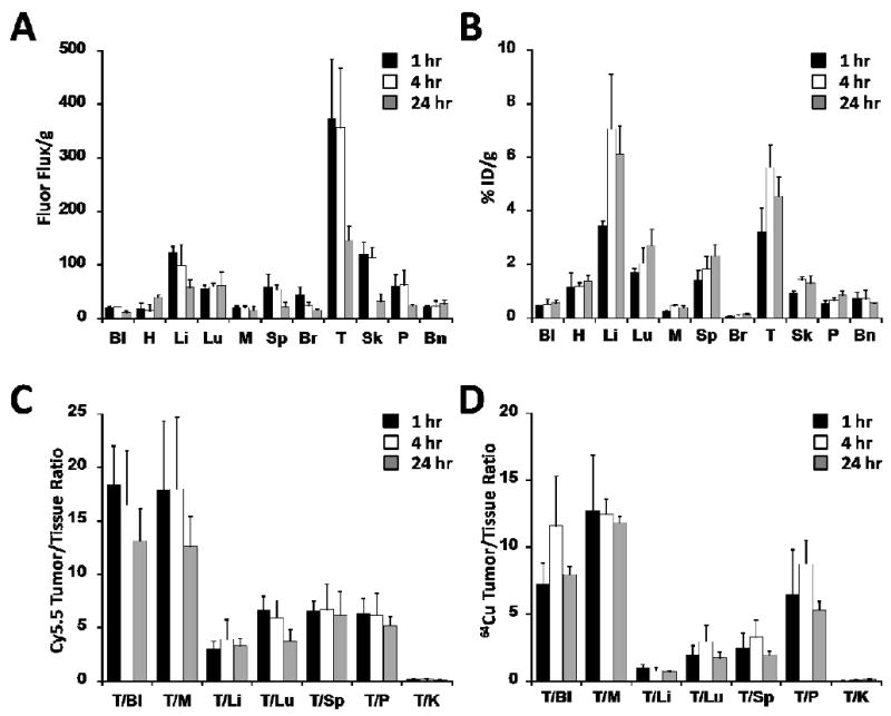
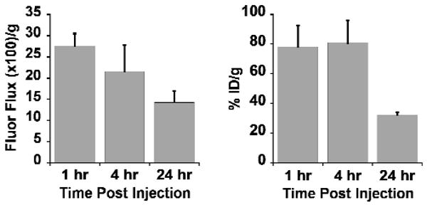
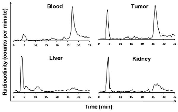
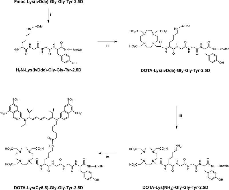
Previously, we used directed evolution to engineer mutants of the Ecballium elaterium trypsin inhibitor (EETI-II) knottin that bind to αvβ3 and αvβ5 integrin receptors with low nanomolar affinity, and showed that Cy5.5- or (64)Cu-DOTA-labeled knottin peptides could be used to image integrin expression in mouse tumor models using near-infrared fluorescence (NIRF) imaging or positron emission tomography (PET). Here, we report the development of a dual-labeled knottin peptide conjugated to both NIRF and PET imaging agents for multimodality imaging in living subjects. We created an orthogonally protected peptide-based linker for stoichiometric coupling of (64)Cu-DOTA and Cy5.5 onto the knottin N-terminus and confirmed that conjugation did not affect binding to αvβ3 and αvβ5 integrins. NIRF and PET imaging studies in tumor xenograft models showed that Cy5.5 conjugation significantly increased kidney uptake and retention compared to the knottin peptide labeled with (64)Cu-DOTA alone. In the tumor, the dual-labeled (64)Cu-DOTA/Cy5.5 knottin peptide showed decreased wash-out leading to significantly better retention (p < 0.05) compared to the (64)Cu-DOTA-labeled knottin peptide. Tumor uptake was significantly reduced (p < 0.05) when the dual-labeled knottin peptide was coinjected with an excess of unlabeled competitor and when tested in a tumor model with lower levels of integrin expression. Finally, plots of tumor-to-background tissue ratios for Cy5.5 versus (64)Cu uptake were well-correlated over several time points post injection, demonstrating pharmacokinetic cross validation of imaging labels. This dual-modality NIRF/PET imaging agent is promising for further development in clinical applications where high sensitivity and high resolution are desired, such as detection of tumors located deep within the body and image-guided surgical resection.


Figures

Figure 1
Figure 1
Competition binding of peptides to surface-immobilized integrins. 125I-labeled echistatin was allowed to compete for binding with 5 nM (black bars) or 50 nM (grey bars) unlabeled peptides to detergent-solubilized integrin receptor subtypes αvβ3, αvβ5, α5β1, and αiibβ3. Unlabeled echistatin, which binds to all four integrins with high affinity, was used as a positive control. Error bars represent the standard deviation of measurements performed in triplicate.
Figure 2
Figure 2
NIRF and microPET imaging of knottin probes in murine human tumor xenograft models. Mice bearing U87MG tumors were injected via tail vein with either (A) 1.5 nmol of DOTA/Cy5.5-2.5D, or (D) ∼ 100 μCi of 64Cu-DOTA/Cy5.5-2.5D. For blocking experiments (B, E), mice were co-injected with an excess (0.5 μmol) of unlabeled c(RGDyK) in addition to labeled knottin peptides. Representative images are shown at 1, 2, 4, and 24 hr post injection. T= tumor; K= kidney; Bd= bladder; L= liver. (C) NIRF imaging, represented as the tumor-to-background tissue (Tumor/Tissue) ratio for Cy5.5-2.5D (white bars), DOTA/Cy5.5-2.5D (black bars), and DOTA/Cy5.5-2.5D plus excess unlabeled c(RGDyK) blocking peptide (grey bars). (F) MicroPET imaging, quantified as the % ID/g of 64Cu-DOTA-2.5D (white bars) 64Cu-DOTA/Cy5.5-2.5D (black bars), and 64Cu-DOTA/Cy5.5-2.5D plus an excess of unlabeled c(RGDyK) blocking peptide (grey bars). Error bars represent the SD of measurements performed on at least three mice. (G) Correlation analysis of average tumor to background tissue ratios for NIRF and microPET images acquired at 1, 2, 4, and 24 hr post injection. Error bars represent the SD of measurements in three mice. (* p < 0.05).
Figure 3
Figure 3
Uptake and imaging in MDA-MB-435 cells and xenograft tumors. (A) Uptake of 64Cu-DOTA/Cy5.5-2.5D in U87MG or MDA-MB-435 cells at 0.5, 1, and 2 hr. Blocking studies, indicated by “block”, were performed in parallel using a large molar excess of c(RGDyK). (B) MicroPET images of mice bearing subcutaneous MDA-MB-435 tumors at 1 hr and 24 hr post injection. Blocking experiments with unlabeled c(RGDyK) were performed to further demonstrate probe specificity.
Figure 4
Figure 4
Comparison of Cy5.5 and 64Cu tissue biodistribution. The amount of (A) DOTA/Cy5.5-2.5D or (B) 64Cu-DOTA/Cy5.5-2.5D accumulation was measured in the blood (Bl), heart (H), liver (Li), lung (Lu), muscle (M), spleen (Sp), brain (Br), tumor (T), skin (Sk), pancreas (P), and bone (Bn). (A) Cy5.5 uptake, measured as the fluorescence flux (photons per second) per gram of tissue. (B) 64Cu uptake, quantified as the %ID/g. Comparison of Tumor/Tissue ratios observed at 1, 4, and 24 hr post injection for (C) DOTA/Cy5.5-2.5D or (D) 64Cu-DOTA/Cy5.5-2.5D.
Figure 5
Figure 5
Comparison of Cy5.5 and 64Cu accumulation in the kidney. Uptake of Cy5.5 resulting from injection of DOTA/Cy5.5-2.5D (left panel) was measured as the fluorescence flux (photons per second) per gram of tissue. Uptake of 64Cu-DOTA/Cy5.5-2.5D (right panel) was quantified as the %ID/g.
Figure 6
Figure 6
In vivo metabolic stability of 64Cu-DOTA/Cy5.5-2.5D. Homogenized tissues were analyzed by radio-HPLC and gamma counting 1 hr post injection. The intact radiotracer elutes at around 27 minutes.
Scheme 1
Scheme 1
Chemical conjugation of DOTA and Cy5.5 to knottin peptide 2.5D. The peptide-based crosslinker Fmoc-Lys(ivDde)-Gly-Gly-Tyr was first synthesized and coupled to the N-terminus of the folded knottin peptide 2.5D using succinimide ester activation chemistry. Next, the Fmoc group was removed from the N-terminus of the crosslinker (i: 10% piperidine/DMF, 0.5 hr, RT), and the resulting compound was reacted with a 5-fold molar excess of an NHS ester-activated DOTA group (ii: DMF/2% DIEA, 0.5 hr, RT) to generate DOTA-Lys(ivDde)-Gly-Gly-Tyr-2.5D. Finally, the ivDde group was removed from the ε-amino group of Lys (iii: 2% hydrazine/DMF, 0.5 hr, RT) and the resulting compound was reacted with Cy5.5 NHS ester (iv: DMF/2% DIEA, 0.5 hr, RT) to give the final product: DOTA-Lys(Cy5.5)-Gly-Gly-Tyr-2.5D, which was used for NIRF imaging experiments, or radiolabeled with 64Cu and used for PET imaging experiments.

Similar articles

Cited by

References

    1. Hynes RO. Integrins: versatility, modulation, and signaling in cell adhesion. Cell. 1992;69:11–25. - PubMed
    1. Brooks PC, Clark RA, Cheresh DA. Requirement of vascular integrin alphav beta3 for angiogenesis. Science. 1994;264:569–571. - PubMed
    1. Friedlander M, Brooks PC, Shaffer RW, Kincaid CM, Varner JA, Cheresh DA. Definition of two angiogenic pathways by distinct alphav integrins. Science. 1995;270:1500–1502. - PubMed
    1. Alghisi GC, Ruegg C. Vascular integrins in tumor angiogenesis: mediators and therapeutic targets. Endothelium. 2006;13:113–135. - PubMed
    1. Mizejewski GJ. Role of integrins in cancer: survey of expression patterns. Proc Soc Exp Biol Med. 1999;222:124–138. - PubMed

Publication types

MeSH terms

Substances