. 2010 Sep;19(9):1375-83.
doi: 10.1089/scd.2009.0258.

Stem cells from human-exfoliated deciduous teeth can differentiate into dopaminergic neuron-like cells

Affiliations

Stem cells from human-exfoliated deciduous teeth can differentiate into dopaminergic neuron-like cells

Jinsong Wang et al. Stem Cells Dev. 2010 Sep.

Abstract

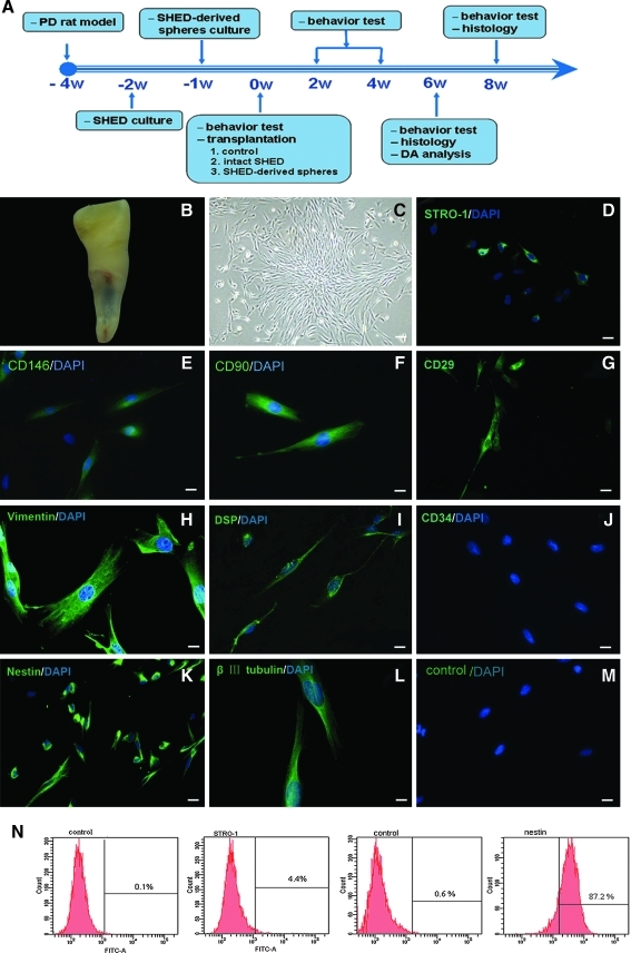
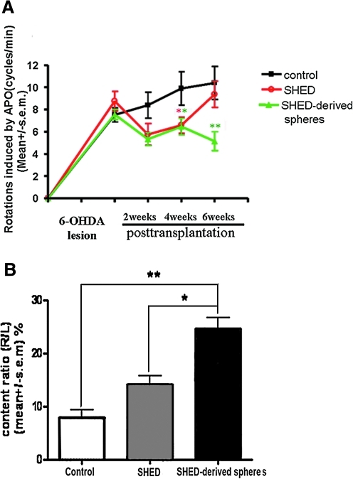
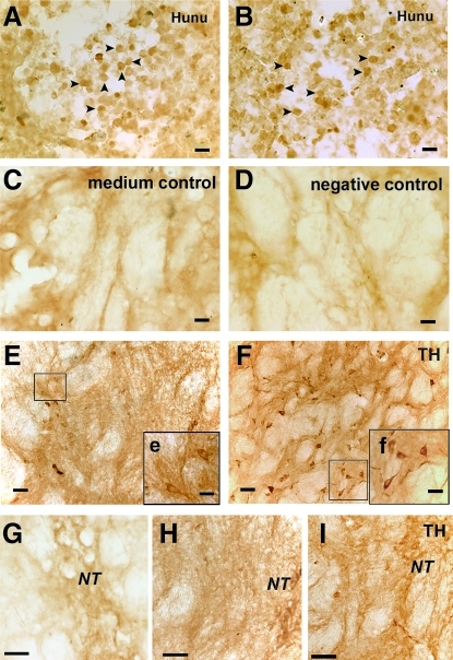
Stem cells from human exfoliated deciduous teeth (SHED) have been identified as a novel population of postnatal stem cells capable of differentiating into neural cells, odontogenic cells, and adipocytes. SHED were reported to differentiate into neural cells based on cellular morphology and the expression of early neuronal markers when cultured under neural inductive conditions. This study therefore investigated the therapeutic efficacy of SHED in alleviating Parkinson's disease (PD) in a rat model. We found that SHED could be induced to form neural-like spheres in a medium optimized for neural stem cells in vitro. After incubation with a cocktail of cytokines including sonic hedgehog, fibroblast growth factor 8, glial cell line-derived neurotrophic factor, and forskolin, these SHED-derived spheres further differentiated into a cell population that contained specific dopaminergic neurons. Moreover, transplantation of SHED spheres into the striatum of parkinsonian rats partially improved the apomorphine-evoked rotation of behavorial disorders compared to transplantation of control SHED. Our data indicate that SHED, potentially derived from neural crest cells, may be an optimal source of postnatal stem cells for PD treatment.


Figures

FIG. 1.
FIG. 1.
Characterization of isolated human-exfoliated deciduous teeth (SHED). (A) Schematic timeline of the procedures conducted for this study. (B) Exfoliated deciduous incisor containing dental pulp. Cultured SHED had a fibroblast-like morphology (C) and expressed STRO-1 (D), CD146 (E), CD90 (F), CD29 (G), vimentin (H), DSP (I), nestin (K), and βIII-tubulin (L), but were negative for CD34 (J), control (M). (N) Flow cytometry analysis of SHED. Numbers in panels represent mean fluorescent intensity of the cells expressing each marker. Scale bars: 20 μm (CE, G, IK, and M) and 10 μm (F, H, and L).
FIG. 2.
FIG. 2.
Differentiation of SHED in vitro. (A) SHED formed sphere-like clusters when cultured in serum-free medium supplemented with epidermal growth factor (EGF) and basic fibroblast growth factor (bFGF). Most cells in the SHED-derived spheres expressed nestin (B) and βIII-tubulin (C), but not TH (D). After incubation with a cytokine cocktail for 7 days, SHED-derived spheres generated neuron-like cells with multiple neurites (E) that expressed βIII-tubulin (F), MAP2 (G), and TH (H). Nuclei were counterstained with DAPI (blue). (I) Expression of TH (68 kDa) was confirmed by western blot analysis (positive control: rat TH [60 kDa]). (J) Real-time PCR revealed that gene transcription levels of the neuronal precursor, nestin, and the early neuronal marker, βIII-tubulin, in differentiated SHED induced from SHED-derived spheres were down-regulated, while expression of the mature neuronal marker MAP2 and TH, the rate-limiting enzyme during DA synthesis, was up-regulated (n = 3). Samples were normalized to the control gene actin. Scale bars: 50 μm (A) and 20 μm (BH). *P < 0.05; **P < 0.01.
FIG. 3.
FIG. 3.
Behavioral and biochemical analysis of parkinsonian rats. (A) Apomorphine-induced rotation in the group of rats transplanted with SHED-derived spheres (n = 12) was ameliorated to a greater extent compared to the negative control group (n = 12), as observed starting 2 weeks after transplantation and then throughout the observation period. This recovery was similar to the intact SHED graft group (n = 12) 2–4 weeks after transplantation, but a better recovery effect was evident in the SHED-derived spheres group. The rats in the control group showed no significant improvement. (B) DA content in the striatum, which reflects the level of active neurotransmitter and its usage efficiency, was measured by high-performance liquid chromatography (HPLC). To avoid variation among individual rats, the result was presented as the ratio of the DA level of the lesion side versus the contralateral (normal) side, which may reflect the actual degree to which synthesis of the neurotransmitter recovered. DA content in the group transplanted with SHED-derived spheres (n = 4) was significantly elevated 6 weeks after transplantation compared to the control group (n = 4) and intact SHED group. *P < 0.05; **P < 0.01.
FIG. 4.
FIG. 4.
Survival and DAergic differentiation of engrafted human cells in parkinsonian rats. Engrafted intact SHED (A) and SHED-derived spheres (B) survived in the striatum of parkinsonian rats 6 weeks post-transplantation and had similar survival rates, as tested by human nuclei staining (Hunu, arrowheads). The group transplanted with basic medium serviced as the medium control in C, and the negative control for anti-human nuclei antibody was shown (D). TH-positive cells with different morphologies were detected around the needle track in the striatum transplanted with intact SHED and SHED-derived spheres. Most of intact SHED possessed fewer and shorter neurites (E, e) and some TH-positive cells of SHED-derived spheres indeed extended more and longer neuritis from the neurosome (F, f), exhibiting typical matured DAergic neurons. More TH-positive fibers (I) were detected around the needle track in the group transplanted with SHED-derived spheres compared to those transplanted with intact SHED grafts (H). (G) Fibers in negative control striatum. Inserts were the higher magnifi cent images. Scale bars: 20 μm (AD), 40 μm (EI), 20 μm (e and f). Abbreviation: Hunu, human nuclei; TH, tyrosine hydroxylase; NT, needle track.

References

    1. Dauer W. Przedborski S. Parkinson's disease: mechanisms and models. Neuron. 2003;39:889–909. - PubMed
    1. Lindvall O. Brundin P. Widner H. Rehncrona S. Gustavii B. Frackowiak R. Leenders KL. Sawle G. Rothwell JC. Marsden CD. Grafts of fetal dopamine neurons survive and improve motor function in Parkinson's disease. Science. 1990;247:574–577. - PubMed
    1. Freed CR. Breeze RE. Rosenberg NL. Schneck SA. Kriek E. Qi JX. Lone T. Zhang YB. Snyder JA. Wells TH. Survival of implanted fetal dopamine cells and neurologic improvement 12 to 46 months after transplantation for Parkinson's disease. N Engl J Med. 1992;327:1549–1555. - PubMed
    1. Olanow CW. Kordower JH. Freeman TB. Fetal nigral transplantation as a therapy for Parkinson's disease. Trends Neurosci. 1996;19:102–109. - PubMed
    1. Greely HT. Hamm T. Johnson R. Price CR. Weingarten R. Raffin T. The ethical use of human fetal tissue in medicine. Stanford University Medical Center Committee on Ethics. N Engl J Med. 1989;320:1093–1096. - PubMed

Publication types

MeSH terms