. 2010 Feb;12(1):43-53.
doi: 10.1089/cell.2009.0043.

Rapid elimination of the histone variant MacroH2A from somatic cell heterochromatin after nuclear transfer

Affiliations

Rapid elimination of the histone variant MacroH2A from somatic cell heterochromatin after nuclear transfer

Ching-Chien Chang et al. Cell Reprogram. 2010 Feb.

Abstract

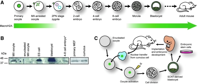
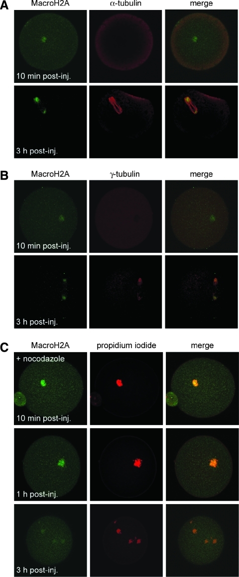
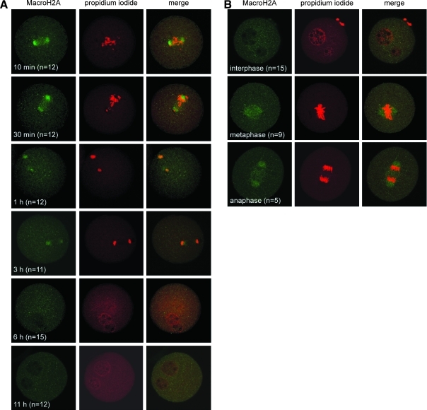
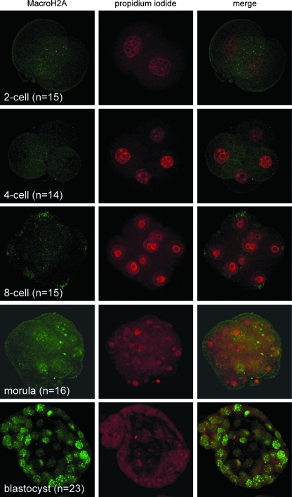
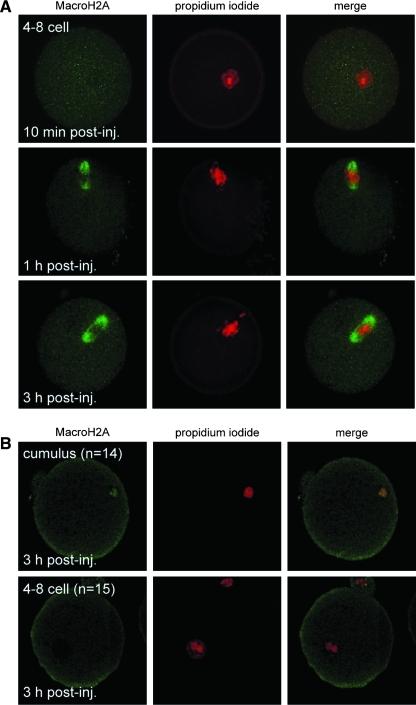
Oocytes contain a maternal store of the histone variant MacroH2A, which is eliminated from zygotes shortly after fertilization. Preimplantation embryos then execute three cell divisions without MacroH2A before the onset of embryonic MacroH2A expression at the 16-cell stage. During subsequent development, MacroH2A is expressed in most cells, where it is assembled into facultative heterochromatin. Because differentiated cells contain heterochromatin rich in MacroH2A, we investigated the fate of MacroH2A during somatic cell nuclear transfer (SCNT). The results show that MacroH2A is rapidly eliminated from the chromosomes of transplanted somatic cell nuclei by a process in which MacroH2A is first stripped from chromosomes, and then degraded. Furthermore, MacroH2A is eliminated from transplanted nuclei by a mechanism requiring intact microtubules and nuclear envelope break down. Preimplantation SCNT embryos express endogenous MacroH2A once they reach the morula stage, similar to the timing observed in embryos produced by natural fertilization. We also show that the ability to reprogram somatic cell heterochromatin by SCNT is tied to the developmental stage of recipient cell cytoplasm because enucleated zygotes fail to support depletion of MacroH2A from transplanted somatic nuclei. Together, the results indicate that nuclear reprogramming by SCNT utilizes the same chromatin remodeling mechanisms that act upon the genome immediately after fertilization.


Figures

FIG. 1.
FIG. 1.
MacroH2A distribution during early mammalian development. (A) MacroH2A (green) is present in the germinal vesicle of primary oocytes and is associated with condensed chromosomes and the first polar body in metaphase II (MII)-arrested oocytes. Upon fertilization, some maternal store MacroH2A is extruded with the second polar body and the remainder is removed prior to formation of the maternal pronucleus (pink). MacroH2A is absent from the paternal pronucleus (blue). Cell division proceeds in the absence of MacroH2A until the morula stage, where MacroH2A expression begins and is sustained throughout the remainder of development. See (Chang et al. 2005) for details. (B) A MacroH2A-specific antibody was used in Western analysis to examine MacroH2A levels in whole-cell lysates from MII oocytes (n = 100), enucleated oocytes (n = 100), embryonic stem (ES) cells (n = 2000), blastocysts (n = 100), and four-cell stage embryos (n = 100) (left panel), as well as mouse embryonic fibroblasts (MEF) (n = 2000) and cumulus cells (n = 2000) (right panel). The asterisk denotes that the faint signal in the last lane may result from residual MacroH2A contained in polar bodies. (C) Summary of experimental design and rationale to investigate the fate of somatic MacroH2A (green nuclei) during somatic cell nuclear transfer (SCNT). A MacroH2A-positive nucleus from a somatic cell—in this case, a cumulus cell—is injected into an enucleated oocyte. Following oocyte activation, successive cell divisions produce a cloned blastocyst, which can be used to obtain embryonic stem cells or can be implanted into a female mouse to produce a reproductive clone. This study investigates the fate of somatic cell MacroH2A dynamics in embryo culture to the blastocyst stage.
FIG. 2.
FIG. 2.
Intracellular distribution of MacroH2A following nuclear transfer. Enucleated metaphase II oocytes (n = 25) were injected with cumulus cell nuclei and observed by indirect immunofluorescence at 10 min (top row), 1 h (middle row), and 3 h (bottom row) postinjection. Injected oocytes were stained with a MacroH2A-specific antibody (left column) and propidium iodide (PI) to visualize DNA (middle column). Green (MacroH2A) and red (PI) signals were merged to show colocalization (right column). Representative images are shown.
FIG. 3.
FIG. 3.
MacroH2A associates with microtubules and aggregates at centrosomes in SCNT oocytes. To investigate intracellular MacroH2A distribution with respect to the spindle apparatus, cumulus cell nucleus-injected oocytes were subjected to coimmunofluorescence using an antibody against MacroH2A and antibodies specific to α-tubulin (A) or γ-tubulin (B) at 10 min (top row) and 3 h (bottom row) postinjection. Green (MacroH2A) and red (α- or γ-tubulin) signals were merged to show colocalization (right column). (C) Treatment of SCNT oocytes with nocodazole prevents the redistribution of MacroH2A (green) from chromosomes (PI) to microtubules.
FIG. 4.
FIG. 4.
MacroH2A distribution in activated SCNT zygotes. (A) Cumulus cell nucleus-injected oocytes were activated with 10 mM SrCl2 and 5 μg/mL Cytochalasin B and observed by indirect immunofluorescence at 10 min, 30 min, 1 h, 3 h, 6 h, and 11 h postactivation. Activated oocytes were stained with a MacroH2A-specific antibody (left column) and propidium iodide (PI) to visualize DNA (middle column). Green (MacroH2A) and red (PI) signals were merged to show colocalization (right column). The number of cells (n) observed at each interval is indicated. (B) Cumulus cell nucleus-injected oocytes were visualized 16 h after activation to examine MacroH2A distribution during the first mitotic division. Indirect immunofluorescence was used to detect MacroH2A during interphase (top row), metaphase (middle row), and anaphase (bottom row). Oocytes were stained with a MacroH2A-specific antibody (left column) and propidium iodide (PI) to visualize DNA (middle column). Green (MacroH2A) and red (PI) signals were merged to show colocalization (right column). The number of cells (n) observed at each interval is indicated.
FIG. 5.
FIG. 5.
Distribution of MacroH2A during preimplantation development of cloned mouse embryos. Embryos formed following cumulus cell nuclear transfer were observed by indirect immunofluorescence at the two-cell (top row), four-cell (second row), eight-cell (third row), morula (fourth row), and blastocyst (fifth row) stages. Cloned embryos were stained with a MacroH2A-specific antibody (left column) and propidium iodide (PI) to visualize DNA (middle column). Green (MacroH2A) and red (PI) signals were merged to show colocalization (right column). The number of embryos (n) observed at each interval is indicated.
FIG. 6.
FIG. 6.
The ability to remodel MacroH2A heterochromatin is dependent on the developmental stage of the embryo. (A) MacroH2A associates with the spindle after injection of a nucleus from a four-cell stage embryo. Injected oocytes were stained 10 min (top row), 1 h (middle row), and 3 h (bottom row) postinjection using a MacroH2A-specific antibody (left column) and propidium iodide (PI) to visualize DNA (middle column). Green (MacroH2A) and red (PI) signals were merged to show colocalization (right column). (B) The enucleated zygote (i.e., both pronuclei have been removed) is incapable of eliminating MacroH2A following nuclear transfer. Enucleated zygotes were injected with nuclei from a cumulus cell (top row) or four-cell stage embryo (bottom row) and were analyzed 3 h postinjection by indirect immunofluorescence with a MacroH2A-specific antibody (left column) and propidium iodide (PI) to visualize DNA (middle column). Green (MacroH2A) and red (PI) signals were merged to show colocalization (right column). The number of zygotes observed (n) at each interval is indicated.

References

    1. Angelov D. Molla A. Perche P.Y., et al. The histone variant macroH2A interferes with transcription factor binding and SWI/SNF nucleosome remodeling. Mol. Cell. 2003;11:1033–1041. - PubMed
    1. Armstrong L. Lako M. Dean W., et al. Epigenetic modification is central to genome reprogramming in somatic cell nuclear transfer. Stem Cells. 2006;24:805–814. - PubMed
    1. Byrne J.A. Simonsson S. Western P.S., et al. Nuclei of adult mammalian somatic cells are directly reprogrammed to oct-4 stem cell gene expression by amphibian oocytes. Curr. Biol. 2003;13:1206–1213. - PubMed
    1. Chadwick B.P. Willard H.F. Cell cycle-dependent localization of macroH2A in chromatin of the inactive X chromosome. J. Cell Biol. 2002;157:1113–1123. - PMC - PubMed
    1. Chakravarthy S. Gundimella S.K. Caron C., et al. Structural characterization of the histone variant macroH2A. Mol. Cell. Biol. 2005;25:7616–7624. - PMC - PubMed

Publication types

LinkOut - more resources