

Towards organ printing: engineering an intra-organ branched vascular tree
- PMID: 20132061
- PMCID: PMC4580374
- DOI: 10.1517/14712590903563352
Towards organ printing: engineering an intra-organ branched vascular tree
Abstract
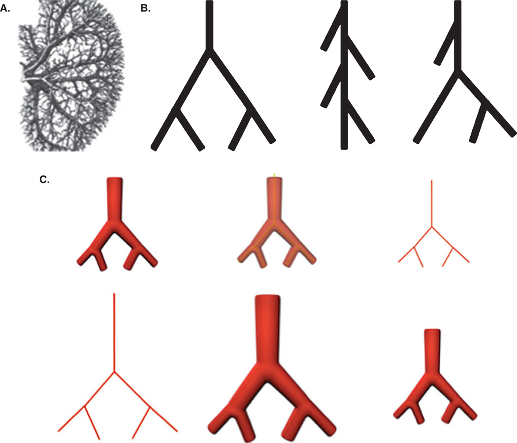
Importance of the field: Effective vascularization of thick three-dimensional engineered tissue constructs is a problem in tissue engineering. As in native organs, a tissue-engineered intra-organ vascular tree must be comprised of a network of hierarchically branched vascular segments. Despite this requirement, current tissue-engineering efforts are still focused predominantly on engineering either large-diameter macrovessels or microvascular networks.
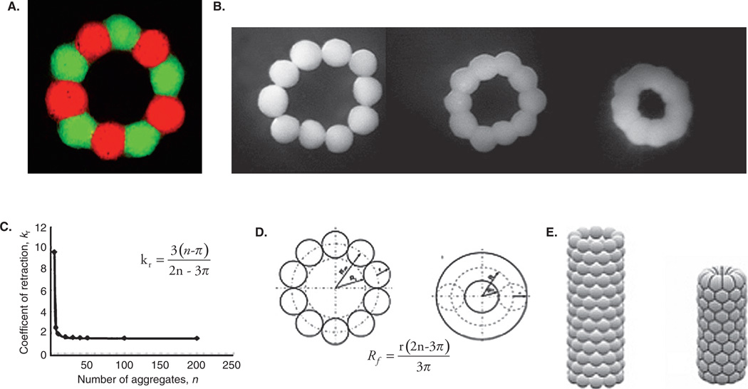
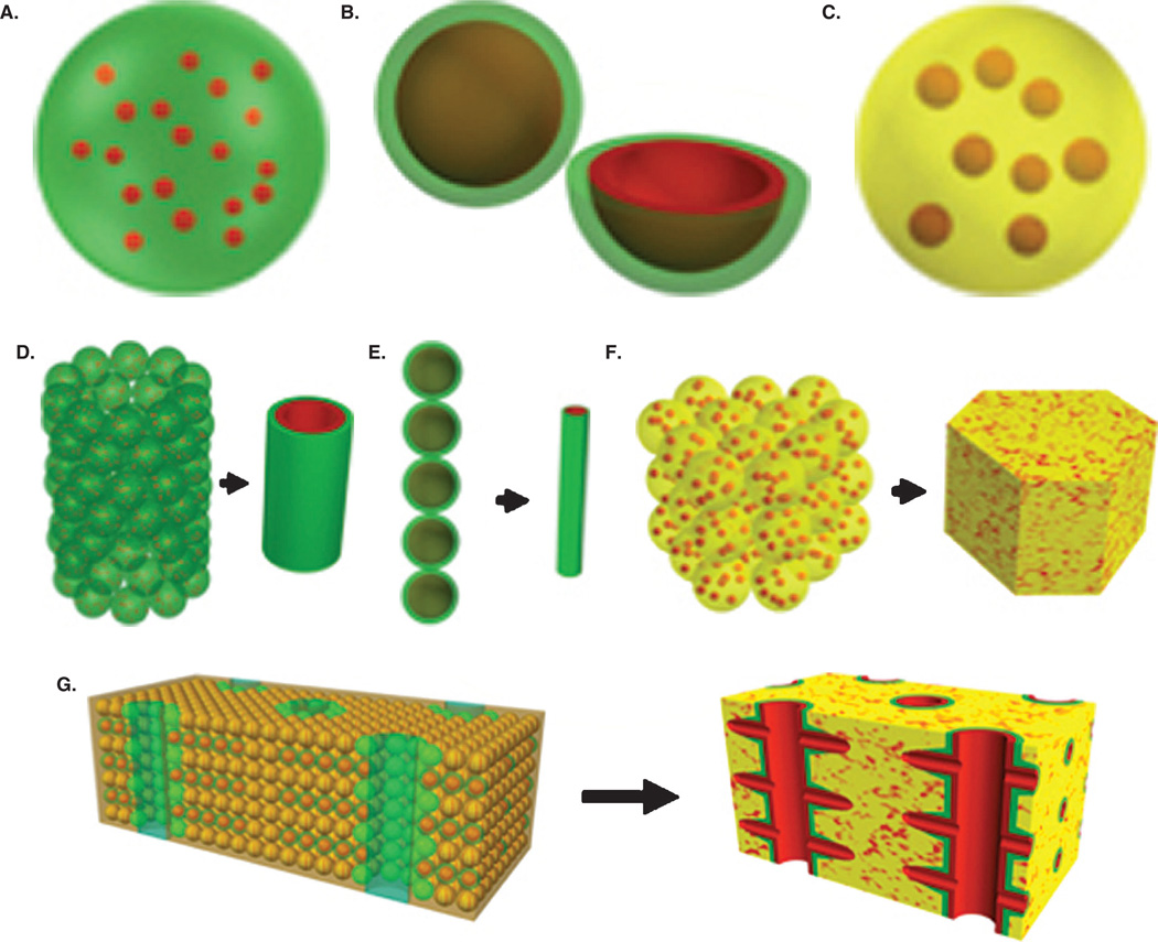
Areas covered in this review: We present the emerging concept of organ printing or robotic additive biofabrication of an intra-organ branched vascular tree, based on the ability of vascular tissue spheroids to undergo self-assembly.
What the reader will gain: The feasibility and challenges of this robotic biofabrication approach to intra-organ vascularization for tissue engineering based on organ-printing technology using self-assembling vascular tissue spheroids including clinically relevantly vascular cell sources are analyzed.
Take home message: It is not possible to engineer 3D thick tissue or organ constructs without effective vascularization. An effective intra-organ vascular system cannot be built by the simple connection of large-diameter vessels and microvessels. Successful engineering of functional human organs suitable for surgical implantation will require concomitant engineering of a 'built in' intra-organ branched vascular system. Organ printing enables biofabrication of human organ constructs with a 'built in' intra-organ branched vascular tree.
Figures
Similar articles
-
Organ printing: tissue spheroids as building blocks.Biomaterials. 2009 Apr;30(12):2164-74. doi: 10.1016/j.biomaterials.2008.12.084. Epub 2009 Jan 26. Biomaterials. 2009. PMID: 19176247 Free PMC article. Review.
-
Bioprinting for vascular and vascularized tissue biofabrication.Acta Biomater. 2017 Mar 15;51:1-20. doi: 10.1016/j.actbio.2017.01.035. Epub 2017 Jan 11. Acta Biomater. 2017. PMID: 28087487 Review.
-
Organ printing: promises and challenges.Regen Med. 2008 Jan;3(1):93-103. doi: 10.2217/17460751.3.1.93. Regen Med. 2008. PMID: 18154465
-
Organ printing: from bioprinter to organ biofabrication line.Curr Opin Biotechnol. 2011 Oct;22(5):667-73. doi: 10.1016/j.copbio.2011.02.006. Epub 2011 Mar 16. Curr Opin Biotechnol. 2011. PMID: 21419621 Review.
-
Biofabrication of small diameter tissue-engineered vascular grafts.Acta Biomater. 2022 Jan 15;138:92-111. doi: 10.1016/j.actbio.2021.11.012. Epub 2021 Nov 13. Acta Biomater. 2022. PMID: 34781026 Review.
Cited by
-
3D Bioprinting for Vascularized Tissue Fabrication.Ann Biomed Eng. 2017 Jan;45(1):132-147. doi: 10.1007/s10439-016-1653-z. Epub 2016 May 26. Ann Biomed Eng. 2017. PMID: 27230253 Free PMC article. Review.
-
Overview of current additive manufacturing technologies and selected applications.Sci Prog. 2012;95(Pt 3):255-82. doi: 10.3184/003685012X13420984463047. Sci Prog. 2012. PMID: 23094325 Free PMC article. Review.
-
Human-scale tissues with patterned vascular networks by additive manufacturing of sacrificial sugar-protein composites.Acta Biomater. 2020 Sep 1;113:339-349. doi: 10.1016/j.actbio.2020.06.012. Epub 2020 Jun 14. Acta Biomater. 2020. PMID: 32553918 Free PMC article.
-
Bioprinting Cellularized Constructs Using a Tissue-specific Hydrogel Bioink.J Vis Exp. 2016 Apr 21;(110):e53606. doi: 10.3791/53606. J Vis Exp. 2016. PMID: 27166839 Free PMC article.
-
Challenges on optimization of 3D-printed bone scaffolds.Biomed Eng Online. 2020 Sep 3;19(1):69. doi: 10.1186/s12938-020-00810-2. Biomed Eng Online. 2020. PMID: 32883300 Free PMC article. Review.
References
-
- Ogawa R, Oki K, Hyakusoku H. Vascular tissue engineering and vascularized 3D tissue regeneration. Regen Med. 2007;2(5):831–837. - PubMed
-
- Lokmic Z, Mitchell GM. Engineering the microcirculation. Tissue Eng Part B Rev. 2008;14(1):87–103. - PubMed
-
- Rivron NC, Liu JJ, Rouwkema J, et al. Engineering vascularised tissues in vitro. Eur Cell Mater. 2008;15:27–40. - PubMed
-
- Rouwkema J, Rivron NC, van Blitterswijk CA. Vascularization in tissue engineering. Trends Biotechnol. 2008;26(8):434–441. - PubMed
-
- Mironov V, Kasyanov V. Emergence of clinical vascular tissue engineering. Lancet. 2009;373(9673):1402–1404. - PubMed
Publication types
MeSH terms
Grants and funding
LinkOut - more resources
Full Text Sources
Other Literature Sources
Research Materials