. 2010 Apr;113(2):374-88.
doi: 10.1111/j.1471-4159.2010.06592.x. Epub 2010 Feb 2.

A novel, high-efficiency cellular model of fibrillar alpha-synuclein inclusions and the examination of mutations that inhibit amyloid formation

Affiliations

A novel, high-efficiency cellular model of fibrillar alpha-synuclein inclusions and the examination of mutations that inhibit amyloid formation

Elisa A Waxman et al. J Neurochem. 2010 Apr.

Abstract

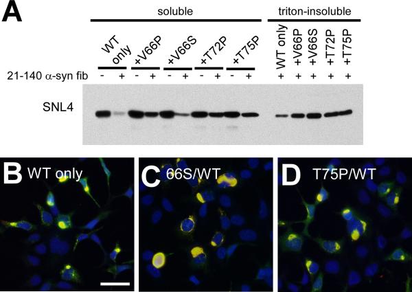
Intracytoplasmic alpha-synuclein (alpha-syn) amyloidogenic inclusions are a major pathological feature of Parkinson's disease, dementia with Lewy body disease and multiple systems atrophy. The mechanisms involved in the formation and inhibition of these aggregates are areas of intense investigation. The present study characterizes a novel cellular model for the study of alpha-syn aggregation, incorporating nucleation-dependent aggregation and a new function for calcium phosphate precipitation. Cultured cells were readily induced to develop large, cytoplasmic alpha-syn filamentous aggregates that were hyperphosphorylated, often ubiquitinated and thioflavin positive. These cellular aggregates formed in the majority of transfected cells and recruited approximately half of endogenously expressed alpha-syn. Using this system, we examined single-point mutations that inhibit alpha-syn amyloid formation in vitro. Three mutations (V66P, T72P and T75P) significantly hindered alpha-syn aggregation in this cell model. The T75P mutant, which could abrogate amyloid formation of wild-type alpha-syn in vitro, did not prevent wild-type alpha-syn cellular aggregates. These studies suggest that the propensity of alpha-syn to form cellular aggregates may be more pronounced than in isolated in vitro studies. This novel high-efficiency cellular model of alpha-syn aggregation is a valuable system that may be used to further understand alpha-syn aggregation and allow for the generation of future therapeutics.


Figures

Figure 1
Figure 1
Double-immunofluorescence with SNL4 (green) and pSer129 (red) on QBI293 cells transfected with WT α-syn in the absence (A) or presence (B) of recombinant, sonicated WT α-syn fibril treatment. α-Syn appeared diffuse in the absence of recombinant fibrils (A). With recombinant fibril treatment (B), a large number of large cellular aggregates were observed (arrows). Faint immunostaining of transfected cells without cellular aggregates was also observed (arrowhead). However, SNL4 also immunolabeled an overabundance of exogenous, recombinant fibrils. pSer129 immunolabeling more specifically recognized intracellular aggregates. (C) Confocal microscopy of double-immunofluorescence of QBI293 cells after transfection and treatment with recombinant fibrils. Immunostaining with thioflavin S (green) and pSer129 (red) showed abundant, large, fibrous intracellular aggregates that were thioflavin positive. Small punctae of thioflavin immunoreactivity was also observed, consistent with exogenous, recombinant fibrils. Bar scale = 50 μM for A,B; 10 μM for C. Representative confocal image is of a single Z-plane of <0.7 μm. (D) Schematic of human α-syn protein and epitopes of anti-α-syn antibodies. Diagram of α-syn shows the 6 imperfect “KTKEGV” repeats (black boxes). Epitopes of anti-α-syn specific antibodies are indicated in relationship to N-terminal (21–140) and C-terminal (1–120) truncated α-syn. Antibodies Syn514 and SNL4 recognize the N-terminus and require amino acids 2–12. Syn211 recognizes amino acids 125–129. pSer129 was raised against a phospho-peptide from amino acids 123–134 and specifically recognizes phosphorylation at residue Ser129.
Figure 2
Figure 2
Confocal microscopy of double-immunofluorescence of QBI293 cells after transfection and seeding with recombinant α-syn fibrils. Cells were transfected with WT α-syn (A,C,D,E,F) or 1–120 α-syn (B) expression plasmids, and then treated with 1 μM of recombinant in vitro generated WT α-syn (B), 1–120 α-syn (A), or 21–140 α-syn (C–F) that had been fibrillized and sonicated prior to addition to the medium (fibrils). Double-immunofluorescence was performed between α-syn-specific antibodies SNL4 and pSer129 (A–D), SNL4 and Syn514 (E), or Syn514 and pSer129 (F). The extracellular addition of fibrils induced the formation of large, fibrous aggregates. Representative images were of cells fixed 48 hr after removal of calcium phosphate. Panel C shows cells containing small aggregates (arrowheads) that are early in formation, where the majority of intracellular α-syn appears soluble and diffuse. Epitope-specificity of antibodies used indicates that the intracellular α-syn aggregates are primarily composed of endogenously expressed α-syn. Representative images are of a single Z-plane of <0.7 μm. Bar scale = 10 μm.
Figure 3
Figure 3
Biochemical cellular fractionation of QBI293 cells after transfection and seeding with recombinant α-syn fibrils. QBI293 cells were transfected with the WT α-syn expression plasmid. After transfection, recombinant 1–120 α-syn or 21–140 α-syn was added to the media. The recombinant protein was added in soluble form, after fibrillization in vitro (fibril mix), or after fibrillization and isolation by centrifugation at 16,000 × g, where the pellet (large fibrils) was isolated from the supernatant (small fibrils/polymers/soluble protein). Western blot analysis of α-syn immunoreactivity was performed after biochemical cellular fractionation isolating soluble from triton-insoluble α-syn, as described in “Materials and Methods.” The first lane at the left shows biochemical fractionation of cells that did not receive any recombinant α-syn treatment. (A) SNL4 immunoreactivity of soluble samples recognized soluble WT α-syn, resulting from cellular transfection. Triton-insoluble protein was analyzed with (B) SNL4, (C) Syn211, and (D) pSer129 antibodies, and α-syn was identified in samples treated with recombinant fibrillized α-syn protein. Migration of α-syn protein and epitope-specific antibodies show that the addition of recombinant fibrillized protein induced aggregation of endogenously expressed α-syn. Arrowheads indicate the immunobands corresponding to full-length WT α-syn (WT) and 1–120 α-syn (1–120).
Figure 4
Figure 4
Biochemical fractionation of QBI293 cells and SH-SY5Y neuroblastoma after recombinant α-syn fibril mix treatment. (A) Representative immunoblot of biochemical cellular fractionation of QBI293 cells over the course of time. Cells were transfected with the WT α-syn expression plasmid and examined with or without recombinant 21–140 α-syn fibril mix treatment (fib). Western blot analyses show an increase in triton-insoluble, phosphorylated α-syn from 24 hours (after calcium phosphate precipitation removal), to 48 hours and was maintained at 72 hours. (B) SH-SY5Y neuroblastoma that were stably transfected with WT α-syn were treated with 1 μM of recombinant 21–140 α-syn fibril mix (fib) either with or without calcium phosphate precipitation (CaPO4) using a control DNA plasmid (pcDNA3.1). Cells were harvested at 48 hours (after calcium phosphate precipitation removal). Representative immunoblot indicated an increase in triton-insoluble, phosphorylated α-syn only with treatment of both CaPO4 and 21–140 α-syn fibril mix.
Figure 5
Figure 5
Double-immunofluorescence of SH-SY5Y neuroblastoma after recombinant α-syn fibril mix treatment. Double-immunofluorescence was performed with SNL4 (green) and pSer129 (red) on SH-SY5Y neuroblastoma that were stably transfected with WT α-syn. Cells were treated with calcium phosphate precipitation (CaPO4) using the control pcDNA3.1 plasmid (A,B,D,E) and/or recombinant 21–140 α-syn fibril mix (fib) (B–E). Removal of CaPO4 (wash), followed by fibril mix treatment was also investigated (D). α-Syn aggregates were formed when cells were treated with recombinant α-syn fibril mix; however, a significant increase in the propensity to form aggregates was observed with concomitant treatment of both CaPO4 and fibrils. (E) Confocal microscopy image of α-syn aggregates in SH-SY5Y neuroblastoma. Bar scale = 100 μm for A–D; 10 μm for E.
Figure 6
Figure 6
α-Syn proteins with mutations in the hydrophobic region inhibit in vitro fibrillization. (A) ThT fluorometry, (B) K114 fluorometry, and (C) quantitative sedimentation analysis of the formation of large polymers and amyloid for WT, V66P, V66S, T72P, or T75P α-syn after 0, 2, 4 and 9 days of incubation under assembly conditions, as described in “Materials and Methods.” All proteins were incubated at 5 mg/ml. Data represent average ± SD. n = 6 for ThT and K114 fluorometry, and n = 3 for sedimentation analysis.
Figure 7
Figure 7
Comparative biochemical fractionation and double-immunofluorescence analyses of QBI293 cells expressing WT α-syn or α-syn containing single-point mutations. (A) Representative immunoblot of biochemical fractionation on cells transfected with plasmids expressing WT, V66P, V66S, T72P, or T75P human α-syn. Treatment with recombinant 21–140 α-syn fibril mix (fib) produced triton-insoluble WT and V66S α-syn protein. Triton-insoluble T75P α-syn was observed in the experiment shown, but was present at low levels, and only present in one set of experiments (n=5, asterisk (*)). Triton-insoluble α-syn was not observed in the absence of fibril mix treatment (data not shown). (B–E) Double-immunofluorescence on QBI293 cells that were transfected with plasmids for the expression of (B) V66P α-syn, (C) T72P α-syn, (D) T75P α-syn, or (E) V66S α-syn. All conditions were treated with recombinant 21–140 α-syn fibril mix. Double-immunofluorescence between SNL4 (green) and pSer129 (red) antibodies shows that cells expressing V66P or T72P α-syn developed small, infrequent pSer129-positive aggregates (arrows). Cells expressing T75P α-syn and treated with recombinant α-syn fibril mix did not present any pSer129-positive aggregates. Microscopic analysis of cells expressing V66S α-syn (E) was indistinguishable from cells expressing WT α-syn. Bar scale = 50 μm.
Figure 8
Figure 8
Effects of α-syn proteins with mutations in the hydrophobic region on the polymerization of WT α-syn in vitro. (A) ThT fluorometry, (B) K114 fluorometry, and (C) quantitative sedimentation analysis of the formation of amyloid and large polymers for recombinant WT α-syn alone or co-incubated with (+) recombinant α-syn that has been mutated at V66P, V66S, T72P, or T75P. All proteins were incubated at 2.5 mg/ml (for a total of 5 mg/ml in conditions containing both WT and mutant protein) for 4 days. Condition indicated as “+T75P (5)” is that of WT α-syn at 2.5 mg/ml incubated with T75P α-syn at 5 mg/ml. Data represent average ± SD. *, p < 0.0001; #, p = 0.0003; ‡, p = 0.003; †, p = 0.002; §, p = 0.03; **, p = 0.01. n=6 for V66P α-syn and T72P α-syn; n=8 for V66S α-syn; n=14 for T75P α-syn K114 and ThT fluorometry; and n=4 for all sedimentation analyses.
Figure 9
Figure 9
Biochemical fractionation and double-immunofluorescence analysis of QBI293 cells co-expressing WT and mutant α-syn and treated with recombinant α-syn fibril mix. (A) Representative immunoblot with antibody SNL4 of biochemically fractionated cells co-transfected with the WT α-syn expression plasmid with empty pcDNA3.1 vector (WT only), or with two plasmids for expression of both WT and V66P α-syn (+V66P), WT and V66S α-syn (+V66S), WT and T72P α-syn (+T72P), or WT and T75P α-syn (+T75P). Treatment with recombinant 21–140 α-syn fibril mix (fib) produced triton-insoluble α-syn in all conditions. Triton-insoluble α-syn was not observed in the absence of fib treatment (data not shown). n = 4. (B–D) Double-immunofluorescence with SNL4 (green) and pSer129 (red) on QBI293 cells that were transfected with two plasmids for expression of (B) WT α-syn and pcDNA3.1 (WT only), (C) WT α-syn and V66S α-syn, or (D) WT α-syn and T75P α-syn. All conditions formed frequent SNL4 and pSer129 positive aggregates (yellow) that were indistinguishable from cells transfected with only the WT α-syn plasmid. Only merged images are shown. Bar scale = 50 μm.

Similar articles

Cited by

References

    1. Ahn KJ, Paik SR, Chung KC, Kim J. Amino acid sequence motifs and mechanistic features of the membrane translocation of alpha-synuclein. J. Neurochem. 2006;97:265–279. - PubMed
    1. Anderson JP, Walker DE, Goldstein JM, et al. Phosphorylation of Ser-129 is the dominant pathological modification of alpha-synuclein in familial and sporadic Lewy body disease. J. Biol. Chem. 2006;281:29739–29752. - PubMed
    1. Ardley HC, Scott GB, Rose SA, Tan NG, Robinson PA. UCH-L1 aggresome formation in response to proteasome impairment indicates a role in inclusion formation in Parkinson's disease. J. Neurochem. 2004;90:379–391. - PubMed
    1. Biere AL, Wood SJ, Wypych J, et al. Parkinson's disease-associated alpha-synuclein is more fibrillogenic than β- and γ-synuclein and cannot cross-seed its homologs. J. Biol. Chem. 2000;275:34574–34579. - PubMed
    1. Bisaglia M, Schievano E, Caporale A, Peggion E, Mammi S. The 11-mer repeats of human alpha-synuclein in vesicle interactions and lipid composition discrimination: a cooperative role. Biopolymers. 2006;84:310–316. - PubMed

Publication types