. 2010 Feb 4:5:7.
doi: 10.1186/1745-6150-5-7.

Origin of the cell nucleus, mitosis and sex: roles of intracellular coevolution

Affiliations

Origin of the cell nucleus, mitosis and sex: roles of intracellular coevolution

Thomas Cavalier-Smith. Biol Direct. .

Abstract

Background: The transition from prokaryotes to eukaryotes was the most radical change in cell organisation since life began, with the largest ever burst of gene duplication and novelty. According to the coevolutionary theory of eukaryote origins, the fundamental innovations were the concerted origins of the endomembrane system and cytoskeleton, subsequently recruited to form the cell nucleus and coevolving mitotic apparatus, with numerous genetic eukaryotic novelties inevitable consequences of this compartmentation and novel DNA segregation mechanism. Physical and mutational mechanisms of origin of the nucleus are seldom considered beyond the long-standing assumption that it involved wrapping pre-existing endomembranes around chromatin. Discussions on the origin of sex typically overlook its association with protozoan entry into dormant walled cysts and the likely simultaneous coevolutionary, not sequential, origin of mitosis and meiosis.

Results: I elucidate nuclear and mitotic coevolution, explaining the origins of dicer and small centromeric RNAs for positionally controlling centromeric heterochromatin, and how 27 major features of the cell nucleus evolved in four logical stages, making both mechanisms and selective advantages explicit: two initial stages (origin of 30 nm chromatin fibres, enabling DNA compaction; and firmer attachment of endomembranes to heterochromatin) protected DNA and nascent RNA from shearing by novel molecular motors mediating vesicle transport, division, and cytoplasmic motility. Then octagonal nuclear pore complexes (NPCs) arguably evolved from COPII coated vesicle proteins trapped in clumps by Ran GTPase-mediated cisternal fusion that generated the fenestrated nuclear envelope, preventing lethal complete cisternal fusion, and allowing passive protein and RNA exchange. Finally, plugging NPC lumens by an FG-nucleoporin meshwork and adopting karyopherins for nucleocytoplasmic exchange conferred compartmentation advantages. These successive changes took place in naked growing cells, probably as indirect consequences of the origin of phagotrophy. The first eukaryote had 1-2 cilia and also walled resting cysts; I outline how encystation may have promoted the origin of meiotic sex. I also explain why many alternative ideas are inadequate.

Conclusion: Nuclear pore complexes are evolutionary chimaeras of endomembrane- and mitosis-related chromatin-associated proteins. The keys to understanding eukaryogenesis are a proper phylogenetic context and understanding organelle coevolution: how innovations in one cell component caused repercussions on others.


Figures
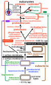
Figure 1
Figure 1
The tree of life and major steps in cell evolution. Archaebacteria are sisters to eukaryotes and, contrary to widespread assumptions, the youngest bacterial phylum [6,13]. This tree topology, coupled with extensive losses of posibacterial properties by the ancestral archaebacterium, explains (without lateral gene transfer) how eukaryotes possess a unique combination of properties now seen in archaebacteria, posibacteria and α-proteobacteria. Eukaryote origins in three stages indicated by asterisks probably immediately followed divergence of archaebacteria and eukaryote precursors from the ancestral neomuran. This ancestor arose from a stem actinobacterial posibacterium by a quantum evolutionary shake-up of bacterial organization - the neomuran revolution [6,12]: surface N-linked glycoproteins replaced murein; ribosomes evolved the signal recognition particle's translational arrest domain; histones replaced DNA gyrase, radically changing DNA replication, repair, and transcription enzymes. The eukaryote depicted is a hypothetical early stage after the origin of nucleus, mitochondrion, cilium, and microtubular skeleton but before distinct anterior and posterior cilia and centriolar and ciliary transformation (anterior cilium young, posterior old: [3]) evolved (probably in the cenancestral eukaryote [9]). Kingdom Chromista was recently expanded to include not only the original groups Heterokonta, Cryptista and Haptophyta, but also Alveolata, Rhizaria and Heliozoa [9], making the name chromalveolates now unnecessary. Excavata now exclude Euglenozoa and comprise just three phyla: the ancestrally aerobic Percolozoa and Loukozoa and the ancestrally anaerobic Metamonada (e.g. Giardia, Trichomonas), which evolved from an aerobic Malawimonas-related loukozoan. Sterols and phosphatidylinositol (PI) probably evolved in the ancestral stem actinobacterium but the ancestral hyperthermophilic archaebacterium lost them when isoprenoid ethers replaced acyl ester lipids.
Figure 2
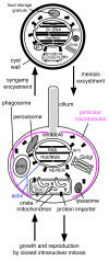
Figure 2
Inferred life cycle and high degree of organellar complexity of the last common ancestor of all extant eukaryotes. This reconstruction assumes that the root of the eukaryotic tree is between Euglenozoa and excavates [7,8]. If so, every homologous character present on both sides of the neokaryote/euglenozoan split must have evolved prior to the cenancestor, provided that its later lateral gene transfer from one to the other can be ruled out, as it can for the complex characters shown. The major uncertainty is whether there were only one centriole and cilium as shown or more likely two of each [9]. In addition to the pellicular microtubules there would also have been centriolar roots consisting of bands of microtubules (probably two if the ancestor was uniciliate and three if biciliate) and a specialized anterior cytostome and cytopharynx for prey ingestion (all not shown for simplicity). The peroxisome (p) was probably attached to the nucleus and the Golgi was probably attached to a centrin body; centrin would also have been associated with the centriole and intranuclearly at mitotic spindle poles. The mitochondrion (m) was probably actually attached to the centriole and/or nucleus. A branched actin cytoskeleton permeating the cytoplasm was linked to nuclear envelope (NE) via KASH/Sun integral membrane protein complexes and to the plasma membrane via membrane-embedded integrin proteins. Syngamy involved fusion of plasma membrane, NE, and probably mitochondria.
Figure 3
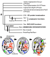
Figure 3
Evolution of eukaryotes from a posibacterium, emphasizing changes in DNA segregation caused by internalization of DNA-membrane attachments. (a). An FtsZ ring between daughter DNA termini (T) divides bacteria; cortical skeletal MreB (blue) and rigid murein wall (brown) control cell shape. (b). Disruptive effects of phagotrophy. Left: flexible glycoproteins (yellow) replaced murein, allowing MreB to become actin (blue) and power phagocytosis, which internalised DNA-membrane attachments (centre); evolution of COP-coated vesicle budding, and fusion with plasma membrane after uncoating, made permanent endomembranes (EM: precursor of ER, NE, Golgi, lysosomes; peroxisomes (P) separated earlier) and disrupted bacterial DNA segregation. (c). Hypothetical origin of simple mitosis in a prekaryote where FtsZ gene duplications evolved stable microtubules and γ-tubulin-containing centromeres still attached to the surface membrane. (d). Accidents in centrosome duplication and phagotrophic membrane internalisation generated a more complex prekaryote II in which stable endomembranes differentiated into peroxisomes (P) and protoendomembranes (EM, i.e. the ancestors of ER and Golgi; see [27]), some associated with the internalised centrosome and DNA; another centrosome remained at the cell surface stabilising it; the actin ring controlled the site of cytokinesis. Ultimately the surface centrosome generated pellicular microtubules and centriolar and ciliary microtubules of the cenancestral eukaryote; the ER-associated MNC nucleated its intranuclear spindle. (e). The first eukaryote. (f). Adding NPCs, mitochondria, and cilium, and nuclear chromosome linearization and kinetochore evolution, made the cenancestral eukaryote, shown in G1 of the cell cycle; bacterial ingestion was via a specialised microtubule-supported pocket-like cytostome (CY) at the apical ciliary end, making the cell asymmetric.
Figure 4
Figure 4
Evolutionary differentiation of endomembranes. (a). Schematic tree for controlling small GTPases [124,128]. Sar-1 and Arf-1 have an extra, derived insertion, so the root cannot be in that branch. Because of disparate rates of evolution among paralogues and the shortness of the molecules it is unclear from trees whether the seven eukaryotic clades (lower) are all mutually related as shown and which of the four bacterial clades (upper) are their closest relatives. (b). After endomembranes, peroxisomes, and plasma membrane became distinct genetic membranes (Fig. 3b) most secretory ribosomes were on old DNA-bearing cisternae; the first COP/adaptin coats generated vesicles (V) from the protoendomembrane/phagosome; early SNAREs (SN, left) fused them with the plasma membrane. Endomembrane differentiation improved digestion by targeting digestive enzymes specifically to phagosomes, mediated by successive concerted duplications and divergence of coat proteins, cognate SNAREs able to bind to them, and associated small GTPases. Primary specialisation between digestion and synthesis involved clathrin vesicles (CL) associated with plasma membrane SNAREs (SNP) and COPII vesicles associated with endomembrane SNAREs (SNE). Interpolation of Golgi, by mutual fusion of uncoated COPII vesicles, stabilised by COPI-mediated recycling (right), allowed specialisation between lysosomes and surface growth. For a fuller discussion of endomembrane origins see [91].
Figure 5
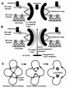
Figure 5
Two-phase origin of the nuclear envelope and trans-envelope transport. (a). Nucleoporins (Nups) forming the octagonal cylindrical scaffold evolved by duplications of coat proteins of COPII secretory vesicles with α-solenoid and/or β-propeller domains, being attached by integral membrane Nups descended from actinobacterial membrane proteins; (b). NPC lumens were narrowed by plugs of FG-repeat-rich Nups, which form a dynamic gel-like meshwork that prevents passive diffusion of macromolecular complexes and mediates active specifically-targeted nucleocytoplasmic exchange by carrier complexes, typically consisting of large karyopherin proteins and their cargo either bound directly or by adaptors. (c). Phase I surface view, showing complete Ran GTPase-mediated fusion of RER cisternae prevented by COPII coat proteins (black blobs) remaining in place to become octagonal NPC scaffolds.
Figure 6
Figure 6
Role of Sun-domain and KASH-domain proteins in nuclear envelope architecture. Sun-domain proteins embedded in the inner membrane attach it directly to the DNA surface of the peripheral heterochromatin (the nucleoskeleton). Their Sun-domains (yellow) bind to the KASH domains (purple) of proteins embedded in the outer membrane, which attach it to the cytoplasmic actin filaments, microtubules, and centrosome of the cytoskeleton. Grey pentagons represent the membrane spanning domain(s) of the KASH-domain proteins and grey rods their flexible cytoskeleton-binding N-terminal domains, which differ greatly among the various types. Microtubules may be attached to KASH-domain proteins either by kinesin or dynein motors. The firm Sun-KASH linker complex (known as LINC) within the perinuclear cisterna holds the inner and outer membrane domains of the NE together with the correct spacing and transmits mechanical forces from cytoskeleton to nucleoskeleton or vice versa without damaging it. Some eukaryotes, e.g. animals, lobose amoebae and peridinean dinoflagellates, (probably polyphyletically) evolved a proteinaceous lamina beneath the inner membrane to further strengthen the nuclear periphery, but this was probably absent in the first eukaryotes; additionally to the universal interactions shown, in animals only cytoplasmic intermediate filaments (IF) attach to KASH proteins via plectin adaptors and lamin IF proteins associate with the intranuclear domain of Sun proteins. For simplicity the fact that Sun-domain proteins are homodimers with a coiled coil domain between their two membrane-spanning and chromatin binding domains (lumped here as grey rectangles) and two Sun domains is not depicted.

Similar articles

Cited by

References

    1. Cavalier-Smith T. A revised six-kingdom system of life. Biol Rev Camb Philos Soc. 1998;73(3):203–266. doi: 10.1017/S0006323198005167. - DOI - PubMed
    1. Stanier RY. In: Organization and Control in Prokaryotic and Eukaryotic Cells (Society for General Microbiology Symposium no 20) Charles HP, Knight JG, editor. Cambridge: Cambridge University Press; 1970. Some aspects of the biology of cells and their possible evolutionary significance; pp. 1–38.
    1. Cavalier-Smith T. The phagotrophic origin of eukaryotes and phylogenetic classification of Protozoa. Int J Syst Evol Microbiol. 2002;52(Pt 2):297–354. - PubMed
    1. Cavalier-Smith T. The origin of eukaryotic and archaebacterial cells. Ann N Y Acad Sci. 1987;503:17–54. doi: 10.1111/j.1749-6632.1987.tb40596.x. - DOI - PubMed
    1. Cavalier-Smith T. Origin of the cell nucleus. BioEssays. 1988;9(2-3):72–78. doi: 10.1002/bies.950090209. - DOI - PubMed

Publication types

Substances

LinkOut - more resources