. 2010 Feb 4:9:30.
doi: 10.1186/1476-4598-9-30.

Prosaposin down-modulation decreases metastatic prostate cancer cell adhesion, migration, and invasion

Affiliations

Prosaposin down-modulation decreases metastatic prostate cancer cell adhesion, migration, and invasion

Siyi Hu et al. Mol Cancer. .

Abstract

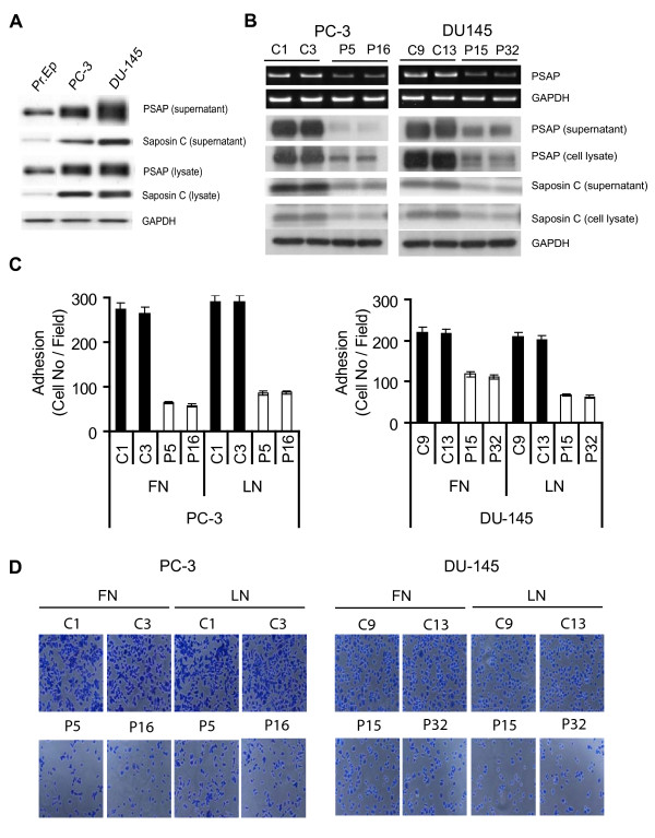
Background: Factors responsible for invasive and metastatic progression of prostate cancer (PCa) remain largely unknown. Previously, we reported cloning of prosaposin (PSAP) and its genomic amplification and/or overexpression in several androgen-independent metastatic PCa cell lines and lymph node metastases. PSAP is the lysosomal precursor of saposins, which serve as activators for lysosomal hydrolases involved in the degradation of ceramide (Cer) and other sphingolipids.

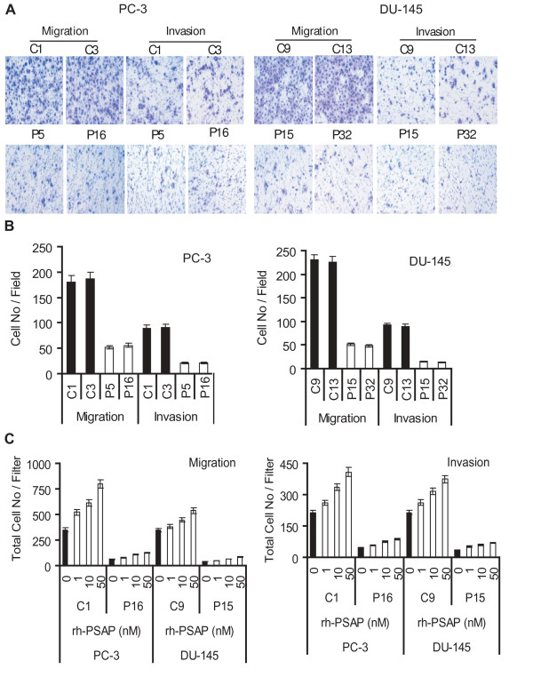
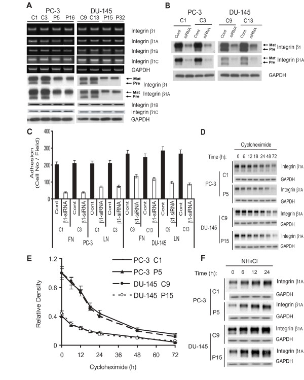
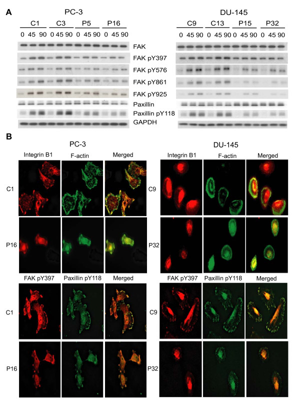
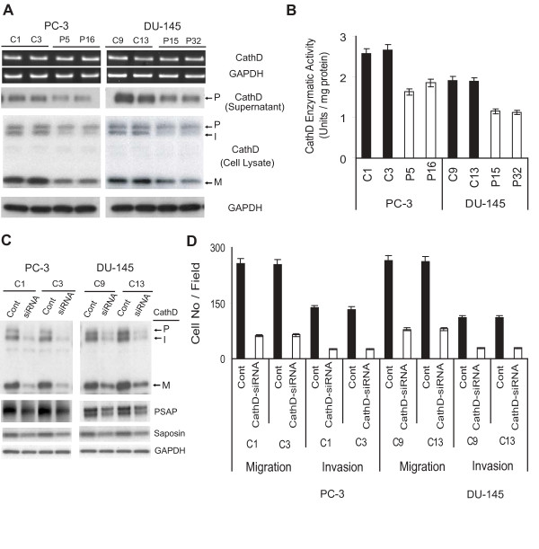
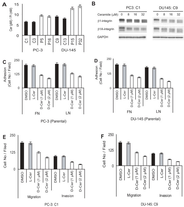
Results: Our current data show that, in metastatic PCa cells, stable down-modulation of PSAP by RNA-interference via a lysosomal proteolysis-dependent pathway decreased beta1A-integrin expression, its cell-surface clustering, and adhesion to basement membrane proteins; led to disassembly of focal adhesion complex; and decreased phosphorylative activity of focal adhesion kinase and its downstream adaptor molecule, paxillin. Cathepsin D (CathD) expression and proteolytic activity, migration, and invasion were also significantly decreased in PSAP knock-down cells. Transient-transfection studies with beta1A integrin- or CathD-siRNA oligos confirmed the cause and effect relationship between PSAP and CathD or PSAP and Cer-beta1A integrin, regulating PCa cell migration and invasion.

Conclusion: Our findings suggest that by a coordinated regulation of Cer levels, CathD and beta1A-integrin expression, and attenuation of "inside-out" integrin-signaling pathway, PSAP is involved in PCa invasion and therefore might be used as a molecular target for PCa therapy.


Figures

Figure 1
Figure 1
PSAP gene silencing decreases metastatic PCa cell adhesion to basement membrane proteins. (A) PSAP over-expression in metastatic prostate cancer cell lines. Equal amount of cell lysates or supernatants from PC-3, DU-145, and normal prostate epithelial (Pr.Ep) cells were subjected to western blotting with anti-PSAP and saposin C antibodies. (B) Stable PSAP down-modulation was accomplished by short-hairpin RNA targeted at PSAP gene. PC-3 and DU-145 metastatic PCa cell lines were stably transfected with a G418-resistant vector containing a shRNA sequence specific for human PSAP or a scrambled control sequence. Total RNA was extracted for RT-PCR (top). GAPDH transcript was used as an internal control for RNA loading. Cell lysates and culture supernatants were subjected to immunoblotting with PSAP antibody. GAPDH antibody was used for protein loading. (C and D) PC-3 and DU-145 cell adhesion to ECM proteins was examined by seeding 1.5 × 10⁴ cells per well in 96-well plates pre-coated with 10 μg/ml fibronectin (FN) or laminin (LN). After 2 h incubation, adhered cells were fixed and stained with toluidine blue. Cells were photographed and counted from ten random fields at 100 × magnification. Columns, mean of three independent samples run together; bars, ± SEM, p < 0.0001, ANOVA was used to compare PSAP-KD and control clones. Each experiment was repeated three times independently. C1 and C3 in PC-3 and C9 and C13 in DU-145 were control clones (shRNA-scrambled vector) and P5 and P16 in PC-3 and P15 and P32 in DU-145 were PSAP-KD clones (shRNA-PSAP).
Figure 2
Figure 2
The PSAP expression correlated with migratory and invasive potential of prostate cancer cell lines. (A) Transwell filter migration and invasion assays. Stable PSAP-KD clones of PC-3 and DU-145 were seeded on transwell filters and incubated for 24 h. Basal medium containing 5% FBS was used as chemo-attractant. For the invasion assay, the membrane was pre-coated with 50 μg Matrigel. (B) Cells migrated or invaded were counted from ten random fields at 100× magnification using a phase-contrast microscope. (C) Effects of rhPSAP on cell migration and invasion. A representative control and PSAP-KD clone from each cell line was seeded on transwell filters and incubated for 24 h (migration) or 48 h (invasion). As a chemo-attractant, basal medium containing 0.5% FBS and various amount of rhPSAP (0, 1, 10, 50 nM) were included in the lower compartment of the transwell filters. Each bar represented the mean ± SEM of three independent experiments, each in quadruplicates. ANOVA was used to examine the significance of the data (p < 0.0001) comparing the PSAP-KD clones relative to control clones in each cell line or among different treatment concentrations for rhPSAP and control.
Figure 3
Figure 3
Effect of PSAP down-modulation on β1A-integrin expression and PCa cell adhesion to FN and LN. (A) PSAP down-modulation reduces β1-integrin expression. Total RNA was extracted for RT-PCR with primers specific for integrin-β1A, -β1B, and -β1C, and GAPDH. Cell lysates were analyzed by western blotting with antibodies against β1-integrin and its isoforms β1A, β1B, and β1C and GAPDH. (B) Transient down modulation of β1-integrin expression. Previously established control stable clones of PC-3 and DU-145 cells were transiently transfected with β1 integrin- or scrambled-siRNA oligos. After 48 h, cell lysates were analyzed for integrin β1 and β1A expression by western blotting. (C) Inhibition of PCa cell adhesion by transient transfection with integrin β1-siRNA. After siRNA transfection, cells were subjected to adhesion assay on FN- or LN-coated 96-well plates as described in "Materials and Methods". (D) β1A-integrin stability. The steady-state β1A protein levels were evaluated by treating cells with cycloheximide (12.5 μg/ml) for up to 72 h and immunoblotting with anti-β1A antibody. (E) The half-live of β1A-integrin was evaluated by densitometric analysis of immunoblotting bands using the Quantity One software and the β1A protein levels were calculated as percentage of non-treatment values after normalization using GAPDH for loading control. (F) Effect of inhibition of lysosomal proteolysis on β1A-integrin expression. Cells were incubated in the presence or absence of the lysosomal proteolysis inhibitor, NH4Cl (50 mM) for up to 24 h. Cell lysates were analyzed for β1A protein expression by immunoblotting. The β1A-integrin degradation curve was calculated as described above. Columns, mean of three independent samples run together; bars, ± SEM, p < 0.0001, Two-sample t-tests with Satterthwaite corrections were used to compare β1-siRNA versus scrambled siRNA oligos transfected cells following adhesion to FN or LN. ANOVA was used to examine the significance of the data (p < 0.05) among different cycloheximide treatment periods in PSAP-KD versus control clones. Similar results were obtained from three independent experiments.
Figure 4
Figure 4
PSAP down-modulation decreased FAK activity and prevented β1A-integrin clustering and proper assembly of focal adhesion complex. (A) PSAP down-modulation reduced phosphorylation of FAK and paxillin. Cells were incubated in suspension with gentle rotation for 45 min and then plated onto FN- or LN-coated dishes for 45 or 90 min. Whole cell lysates were extracted and equal amount of proteins were used for immunoprecipitation with anti-FAK or-paxillin antibody and immunoblotting with phospho-specific antibodies against Tyr-397, -576, -861, -925 of FAK or Tyr-118 of paxillin. (B) Effect of PSAP down-modulation on β1A-integrin clustering and focal adhesion complex assembly. Cells were plated onto FN- or LN-coated slides for 2 h, fixed and permeabilized. Immunofluorescence staining was performed with primary antibodies against integrin β1A, FAK pY397 and paxillin pY118 followed by Cy3 (red) or FITC (green)-conjugated secondary antibodies. F-actin was stained by Oregon Green 488-phalloidin (green). All images were taken by a Leica DM RA2 fluorescence microscope. Consistent data were obtained from three independent experiments.
Figure 5
Figure 5
Down-regulation of cathepsin D expression and activity decreased migration and invasion in PSAP-KD cells. (A) PSAP down-modulation reduced CathD expression and activity. Total RNA was subjected to RT-PCR using specific primers for CathD. Cell lysates and culture supernatants were prepared from parallel dishes and analyzed by immunoblotting with an anti-CathD monoclonal antibody which recognizes proCathD (P), intermediate CathD (I), and mature CathD (M). (B) Whole cell extracts were also assayed for CathD enzymatic activity using a kit with a fluorimetric substrate. The enzymatic activity of CathD was calculated as units/mg total protein. Columns, mean of three independent samples run together; bars, ± SEM. ANOVA was used to examine the significance of the data (p < 0.0001) comparing PSAP-KD clones versus control clones. (C) Transient down-modulation of CathD and its effect on PSAP expression. Control clones of PC-3 and DU-145 cell lines were transiently transfected with specific CathD- or control-siRNA oligos. After 48 h, cell lysates were analyzed for CathD, PSAP, and saposin C expression by immunoblotting. (D) Migration and invasion assays were performed on parallel-transfected tissue culture plates as described in the legend to Fig. 2. Columns, mean of three independent samples run together; bars, ± SEM, p < 0.0001, Two-sample t-tests with Satterthwaite corrections were used to compare CathD-siRNA versus scrambled-siRNA oligos transfected cells. Similar results were obtained from three independent experiments.
Figure 6
Figure 6
Effect of ceramide on PCa cell adhesion, migration, and invasion. (A) PSAP down-modulation increased Cer levels in PSAP-KD cells. PSAP-KD and control clones of PC-3 and DU-145 cells were subjected to matrix-assisted laser desorption mass spectrometric analysis as described in "Materials and Methods". The assay was performed in duplicate and repeated twice independently. Cer content was quantified and calibrated to the intracellular phosphate (Pi) level and depicted as Cer (pM)/Pi (nM). (B) Effect of Cer on β1A-integrin expression. Control clones of PC-3 and DU-145 cells were treated with active or inactive Cer analogs or vehicle (DMSO) at the indicated concentrations for 36 h. Cell lysates were subjected to immunoblotting with specific antibodies against total β1-integrin or β1A-isoform. Inactive Cer analog and DMSO did not affect integrins expression level. GAPDH antibody was used as control loading. (C and D) Effect of Cer on PCa cell adhesion. Parental PC-3 and DU-145 cells were treated with cell permeable natural Cer analog (C6-D-e-Cer; D-Cer), inactive Cer (C6-L-e-Cer; L-Cer), or vehicle (DMSO) at 1 or 2 μM for three to five days and then, subjected to cell adhesion on FN- or LN-coated plates as described in the legend to Fig. 1. (E and F) Effect of Cer on PCa cell migration and invasion. The effect of Cer on migration and invasion of control PC-3 and DU-145 transfectants was examined by treating cells with active or inactive Cer analogs (described above) followed by migration and invasion assay as explained in the legend to Fig. 2. Each bar represents the mean ± SEM of three independent experiments. ANOVA models with Dunnett and Tukey corrections were used to compare cells treated with DMSO, L-Cer, or D-Cer. Statistically significant differences were set at p < 0.05. Consistent data were obtained from three independent experiments.

Similar articles

Cited by

References

    1. Kishimoto Y, Hiraiwa M, O'Brien JS. Saposins: Structure, function, distribution and molecular genetics. J Lipid Res. 1992;33:1255–1267. - PubMed
    1. Koochekpour S. PSAP (Prosaposin (variant Gaucher disease and variant metachromatic leukodystrophy)) Atlas Genet Cytogenet Oncol Haematol. 2006. http://AtlasGeneticsOncology.org/Genes/PSAPID42980ch10q22.html
    1. Schuette CG, Pierstorff B, Huettler S, Sandhoff K. Sphingolipid activator proteins: proteins with complex functions in lipid degradation and skin biogenesis. Glycobiol. 2001;11:81R–90R. doi: 10.1093/glycob/11.6.81R. - DOI - PubMed
    1. Gopalakrishnan MM, Grosch HW, Locatelli-Hoops S, Werth N, Smolenov E, Nettersheim M, Sandhoff K, Hasilik A. Purified recombinant human prosaposin forms oligomers that bind procathepsin D and affect its autoactivation. Biochem J. 2004;383:507–515. doi: 10.1042/BJ20040175. - DOI - PMC - PubMed
    1. Hiraiwa M, Martin BM, Kishimoto Y, Conner GE, Tsuj S, O'Brien JS. Lysosomal proteolysis of prosaposin, the precursor of saposins (aphingolipid activator proteins): its mechanism and inhibition by ganglioside. Arch Biochem Biophys. 1997;341:17–24. doi: 10.1006/abbi.1997.9958. - DOI - PubMed

Publication types

MeSH terms