. 2010 Feb 4:10:32.
doi: 10.1186/1471-2407-10-32.

Caspase 8 and maspin are downregulated in breast cancer cells due to CpG site promoter methylation

Affiliations

Caspase 8 and maspin are downregulated in breast cancer cells due to CpG site promoter methylation

Yanyuan Wu et al. BMC Cancer. .

Abstract

Background: Epigenetic changes associated with promoter DNA methylation results in silencing of several tumor suppressor genes that lead to increased risk for tumor formation and for progression of the cancer.

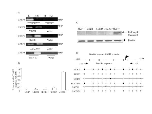
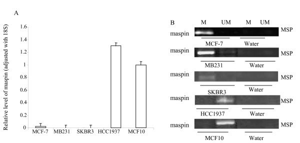
Methods: Methylation specific PCR (MSP) and bisulfite sequencing were used for determination of proapoptotic gene Caspase 8 (CASP8) and the tumor suppressor gene maspin promoter methylation in four breast cancer and two non-tumorigenic breast cell lines. Involvement of histone H3 methylation in those cell lines were examined by CHIP assay.

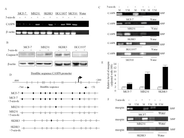
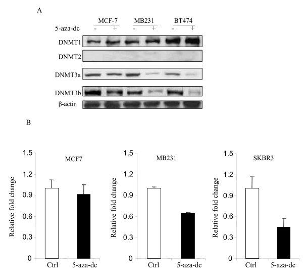
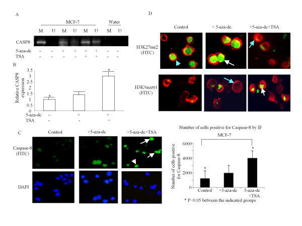
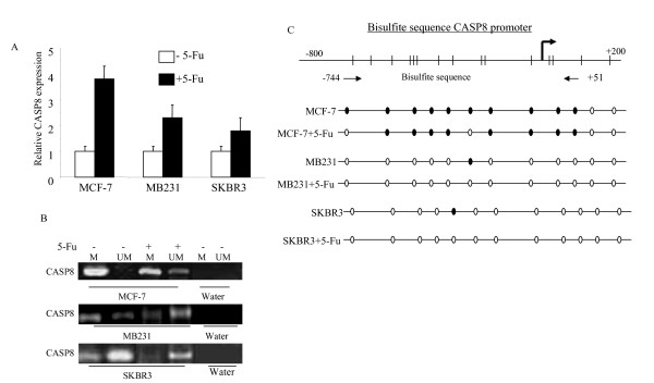
Results: The CpG sites in the promoter region of CASP8 and maspin were methylated in all four breast cancer cell lines but not in two non-tumorigenic breast cell lines. Demethylation agent 5-aza-2'-deoxycytidine (5-aza-dc) selectively inhibits DNA methyltransferases, DNMT3a and DNMT3b, and restored CASP8 and maspin gene expression in breast cancer cells. 5-aza-dc also reduced histone H3k9me2 occupancy on CASP8 promoter in SKBR3cells, but not in MCF-7 cells. Combination of histone deacetylase inhibitor Trichostatin A (TSA) and 5-aza-dc significant decrease in nuclear expression of Di-methyl histone H3-Lys27 and slight increase in acetyl histone H3-Lys9 in MCF-7 cells. CASP8 mRNA and protein level in MCF-7 cells were increased by the 5-aza-dc in combination with TSA. Data from our study also demonstrated that treatment with 5-FU caused a significant increase in unmethylated CASP8 and in CASP8 mRNA in all 3 cancer lines.

Conclusions: CASP8 and maspin expression were reduced in breast cancer cells due to promoter methylation. Selective application of demethylating agents could offer novel therapeutic opportunities in breast cancer.


Figures

Figure 1
Figure 1
CASP8 promoter methylation resulted in decreased mRNA and protein expression. (A) DNA from the indicated cell lines were modified by bisulfite treatment, and MSP were performed with methylated and unmethylated CASP8 PCR primers as described in Methods. The bands detected by methylated primer represented methylated CASP8 (M), and the bands detected by unmethylated primer represented unmethylated CASP8 (UM). (B) RNA were isolated from cells and performed for RT and real-time PCR with CASP8. The relative level of CASP8 gene was normalized to 18 s mRNA as described in Methods. Each bar represented the mean of three independent amplifications with standard deviation (SD). (C) Total protein was isolated from the indicated cells, and Western blot analysis was performed with antibodies specific for full length caspase-8. β-actin was used as loading control. (D). Bisulfite modified DNA from the indicated cells were amplified with CASP8 primer designed according to the bisulfite modified sequence of CpG sites in CASP8 promoter as described in Methods. The amplified PCR products were purified and sequenced to confirm the location of methylated CpG sites. The dark dots represent location of methylated CpG sites in the promoter, and open dots indicate unmethylated CpG sites.
Figure 2
Figure 2
Maspin promoter methylation resulted in decreased mRNA and protein expression. (A) RNA were isolated from cells and performed for RT and real-time PCR with maspin primers. The relative level of the maspin gene was normalized to 18 s as described in Methods. Each bar represented the mean of three independent amplifications with standard deviation (SD). (B) DNA from the indicated cell lines were modified by bisulfite treatment, and MSP were performed with methylated and unmethylated maspin PCR primers as described in Methods. The bands detected by methylated primer represented methylated maspin (M), and the bands detected by unmethylated primer represented unmethylated maspin (UM).
Figure 3
Figure 3
Effect of 5-aza-dc treatment on mRNA, protein expression and promoter methylation of CASP8 and maspin. Cells were treated with 5 μM 5-aza-dc for 3 days. (A) RNA were isolated from the treated and untreated cells and performed for RT-PCR with CASP8 primers. β-actin was used as loading control. (B) Total protein was isolated after cells were treated or untreated with 5-aza-dc. Western blot analysis was performed with antibodies specific for full length caspase-8, and β-actin was used as loading control. (C) DNA were isolated and modified by bisulfite treatment. MSP was performed with methylated and unmethylated CASP8 PCR primers as described in Methods. The bands detected by methylated primer represented methylated maspin (M), and the bands detected by unmethylated primer represented unmethylated maspin (UM). (D) The DNA from 5-aza-dc treated and untreated cells were modified by bisulfite treatment and amplified with CASP8 primer designed according to the bisulfite modified sequence of CpG sites in CASP8 promoter as described in Methods. The amplified PCR products were purified and sequenced for confirmation the CpG sites methylation. The dark dots represented methylated CpG sites in the promoter, and open dots indicated unmethylated CpG sites. (E) RNA from 5-aza-dc treated and untreated cells were used to perform RT and real-time PCR with maspin primers. The relative level of maspin gene was normalized to 18 s as described in Methods. Each bar represented the mean of three independent amplifications with standard deviation (SD). (F) DNA from cells treated and untreated with 5-aza-dc were modified by bisulfite treatment, and MSP was performed with methylated and unmethylated maspin PCR primers as described in Methods. The bands detected by methylated primer represented methylated maspin (M), and the bands detected by unmethylated primer represented unmethylated maspin (UM).
Figure 4
Figure 4
Effect of 5-aza-dc on DNMTs, and H3K9me2. (A) Total protein from the 5-aza-dc treated and untreated cells was used to perform Western blot analysis with antibodies specific against DNMT1, DNMT2, DNMT3a and DNMT3b as described in Methods. Antibody against β-actin was also used as loading control. (B) Chromatins were extracted from cells with or without 5-aza-dc treatment as described in Methods. Chromatin immunoprecipitation was performed with antibody specific against histone H3K9me2, and recovered DNA fragments were used for Q-PCR with promoter specific primers of CASP8. Fold change of H3K9me2 occupancy was calculated based on the control (without 5-aza-dc treatment). Each bar represented the mean of three independent amplifications with standard deviation (SD).
Figure 5
Figure 5
Effect of TSA in combination with 5-aza-dc on CASP8. (A) DNA was isolated from the TSA and 5-aza-dc treated and untreated cells and MSP was performed with methylated and unmethylated CASP8 PCR primers as described in Methods. The bands detected by methylated primer represented methylated CASP8 (M), and the bands detected by unmethylated primer represented unmethylated CASP8 (UM). (B) RNA was isolated from the TSA and 5-aza-dc treated and untreated cells. RT and Q-PCR was performed with CASP8 primers. The relative level of CASP8 mRNA was normalized to 18 s as described in Methods. Each bar represented the mean of three independent amplifications with standard deviation (SD), *p < 0.05 between indicated groups. (C) Immunofluorescence analysis was performed with cleaved caspase-8 antibody followed by incubation with anti-goat IgG-FITC and mounted with DAPI mounting medium (left panel). The arrows indicate the FITC labeled cleaved caspase-8 (green in the top panel of left), and the cell nucleus was labeled by DAPI (blue in bottom panel of left). The cells with positive staining for cleaved caspase-8 were counted in five different areas and adjusted with total number of cells (right panel). Each bar represents the mean of number of cells positive for cleaved caspase-8 in the five areas with standard deviation (SD), * p < 0.05 between the indicated groups. (D) Double immunofluorescence analysis was performed with Di-Methyl Histone H3 (Lys27) or Acetyl Histone H3 (Lys9) antibodies followed by actin antibody as described in Methods. The nucleus positive stained with Di-Methyl Histone H3 (Lys27) or Acetyl Histone H3 (Lys9) were labeled with FITC as green (indicated by white arrows), and the cytoplasm stained with actin was labeled with Tex-red as red (indicated with light blue arrows).
Figure 6
Figure 6
Effect of 5-Fu on CASP8. (A) MCF-7, MB231, and SKBR3 cells were treated with or without 5-Fu (10 μM) for 3 days. RNA was isolated from the treated and untreated cells and RT-real-time PCR was performed with CASP8 and 18 s primers. Each bar represented the mean relative level of CASP8 and standard deviation (SD) from three measurements and normalized to 18 s. (B) DNA was isolated from the 5-Fu treated and untreated cells and modified with bisulfite treatment. MSP was performed with methylated and unmethylated CASP8 primers. The bands detected with methylated primer indicated methylated CASP8 (M), and the bands from unmethylated primer indicated unmethylated CASP8 (UM). (C) Bisulfite modified DNA from the indicated 5-Fu treated, and untreated cells were amplified with CASP8 primer designed according to the bisulfite modified sequence of CpG sites in CASP8 promoter as described in Methods. The amplified PCR products were purified and sequenced for confirmation of the CpG sites methylation. The dark dots in the bottom panel represented methylated CpG sites in the promoter, and open dots indicated unmethylated CpG sites.

Similar articles

Cited by

References

    1. Chiang PK, Gordon RK, Tal J, Zeng GC, Doctor BP, Pardhasaradhi K, McCann PP. S-Adenosylmethionine and methylation. FASEB J. 1996;10:471–480. - PubMed
    1. Baylin SB, Herman JG, Graff JR, Vertino PM, Issa JP. Alterations in DNA methylation: a fundamental aspect of neoplasia. Adv Cancer Res. 1998;72:141–196. full_text. - PubMed
    1. Yang X, Yan L, Davidson NE. DNA methylation in breast cancer. Endocr Relat Cancer. 2001;8:115–127. doi: 10.1677/erc.0.0080115. - DOI - PubMed
    1. Widschwendter M, Jones PA. DNA methylation and breast carcinogenesis. Oncogene. 2002;21:5462–5482. doi: 10.1038/sj.onc.1205606. - DOI - PubMed
    1. Miyakura Y, Sugano K, Konishi F, Ichikawa A, Maekawa M, Shitoh K, Shitoh K, Igarashi S, Kotake K, Koyama Y, Nagai H. Extensive methylation of hMLH1 promoter region predominates in proximal colon cancer with microsatellite instability. Gastroenterology. 2001;121:1300–1309. doi: 10.1053/gast.2001.29616. - DOI - PubMed

Publication types