

A case report of pseudoprogression followed by complete remission after proton-beam irradiation for a low-grade glioma in a teenager: the value of dynamic contrast-enhanced MRI
- PMID: 20132555
- PMCID: PMC2829589
- DOI: 10.1186/1748-717X-5-9
A case report of pseudoprogression followed by complete remission after proton-beam irradiation for a low-grade glioma in a teenager: the value of dynamic contrast-enhanced MRI
Abstract
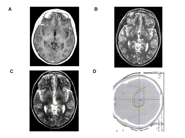
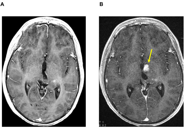
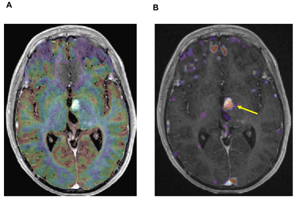
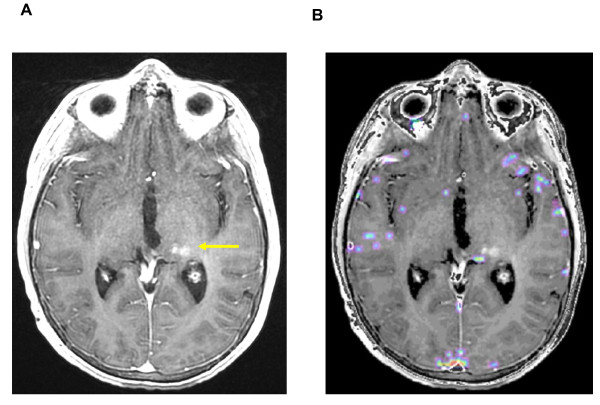
A fourteen years-old boy was treated post-operatively with proton therapy for a recurrent low-grade oligodendroglioma located in the tectal region. Six months after the end of irradiation (RT), a new enhancing lesion appeared within the radiation fields. To differentiate disease progression from radiation-induced changes, dynamic susceptibility contrast-enhanced (DSCE) MRI was used with a T2* sequence to study perfusion and permeability characteristics simultaneously. Typically, the lesion showed hypoperfusion and hyperpermeability compared to the controlateral normal brain. Without additional treatment but a short course of steroids, the image disappeared over a six months period allowing us to conclude for a pseudo-progression. The patient is alive in complete remission more than 2 years post-RT.
Figures
Similar articles
-
Glioma recurrence versus radiation necrosis? A pilot comparison of arterial spin-labeled, dynamic susceptibility contrast enhanced MRI, and FDG-PET imaging.Acad Radiol. 2010 Mar;17(3):282-90. doi: 10.1016/j.acra.2009.10.024. Epub 2010 Jan 12. Acad Radiol. 2010. PMID: 20060750
-
Treatment-related change versus tumor recurrence in high-grade gliomas: a diagnostic conundrum--use of dynamic susceptibility contrast-enhanced (DSC) perfusion MRI.AJR Am J Roentgenol. 2012 Jan;198(1):19-26. doi: 10.2214/AJR.11.7417. AJR Am J Roentgenol. 2012. PMID: 22194475 Review.
-
Differentiation between radiation-induced brain injury and glioma recurrence using 3D pCASL and dynamic susceptibility contrast-enhanced perfusion-weighted imaging.Radiother Oncol. 2018 Oct;129(1):68-74. doi: 10.1016/j.radonc.2018.01.009. Epub 2018 Feb 2. Radiother Oncol. 2018. PMID: 29398151
-
Incidence of pseudoprogression in low-grade gliomas treated with radiotherapy.Neuro Oncol. 2017 May 1;19(5):719-725. doi: 10.1093/neuonc/now194. Neuro Oncol. 2017. PMID: 28453748 Free PMC article.
-
Advanced magnetic resonance imaging methods for planning and monitoring radiation therapy in patients with high-grade glioma.Semin Radiat Oncol. 2014 Oct;24(4):248-58. doi: 10.1016/j.semradonc.2014.06.008. Epub 2014 Jul 26. Semin Radiat Oncol. 2014. PMID: 25219809 Free PMC article. Review.
Cited by
-
Dual contrast perfusion MRI in a single imaging session for assessment of pediatric brain tumors.J Neurooncol. 2012 Aug;109(1):105-14. doi: 10.1007/s11060-012-0872-x. Epub 2012 Apr 19. J Neurooncol. 2012. PMID: 22528798 Free PMC article.
-
MR perfusion and diffusion imaging in the follow-up of recurrent glioblastoma treated with dendritic cell immunotherapy: a pilot study.Neuroradiology. 2011 Oct;53(10):721-31. doi: 10.1007/s00234-010-0802-6. Epub 2010 Nov 25. Neuroradiology. 2011. PMID: 21107549
-
First experiences in treatment of low-grade glioma grade I and II with proton therapy.Radiat Oncol. 2012 Nov 9;7:189. doi: 10.1186/1748-717X-7-189. Radiat Oncol. 2012. PMID: 23140402 Free PMC article.
-
Radiotherapy of high-grade gliomas: current standards and new concepts, innovations in imaging and radiotherapy, and new therapeutic approaches.Chin J Cancer. 2014 Jan;33(1):16-24. doi: 10.5732/cjc.013.10217. Chin J Cancer. 2014. PMID: 24384237 Free PMC article. Review.
-
Re-irradiation of recurrent pediatric ependymoma: modalities and outcomes: a twenty-year survey.Springerplus. 2016 Jun 24;5(1):879. doi: 10.1186/s40064-016-2562-1. eCollection 2016. Springerplus. 2016. PMID: 27386327 Free PMC article.
References
-
- Bakardjiev AI, Barnes PD, Goumnerova LC, Black PM, Scott RM, Pomeroy SL, Billet A, Loeffler JS, Tarbell NJ. Magnetic resonance imaging changes after stereotactic radiation therapy for childhood low grade astrocytoma. Cancer. 1996;78:864–873. doi: 10.1002/(SICI)1097-0142(19960815)78:4<864::AID-CNCR25>3.0.CO;2-S. - DOI - PubMed
-
- Sugahara T, Korogi Y, Tomiguchi S, Shigematsu Y, Ikushima I, Kira T, Liang L, Ushio Y, Takahashi M. Posttherapeutic intra-axial brain tumor: the value of perfusion-sensitive contrast-enhanced MR imaging for differentiating tumor recurrence from non-neoplastic contrast enhancing tissue. AJNR Am J Neuroradiol. 2000;21:901–909. - PMC - PubMed
-
- De Wit, de Bruin HG, Eljkenboom W, Sillevis Smitt PA, Bent MJ van den. Immediate post-radiotherapy changes in malignant glioma can mimic tumor progression. Neurology. 2004;63:535–537. - PubMed
Publication types
MeSH terms
Substances
LinkOut - more resources
Full Text Sources
Medical
