. 2010 May;1798(5):861-71.
doi: 10.1016/j.bbamem.2010.01.018. Epub 2010 Feb 2.

Interaction of the exported malaria protein Pf332 with the red blood cell membrane skeleton

Affiliations

Interaction of the exported malaria protein Pf332 with the red blood cell membrane skeleton

Karena L Waller et al. Biochim Biophys Acta. 2010 May.

Abstract

Intra-erythrocytic Plasmodium falciparum malaria parasites synthesize and export numerous proteins into the red blood cell (RBC) cytosol, where some bind to the RBC membrane skeleton. These interactions are responsible for the altered antigenic, morphological and functional properties of parasite-infected red blood cells (IRBCs). Plasmodium falciparum protein 332 (Pf332) is a large parasite protein that associates with the membrane skeleton and who's function has recently been elucidated. Using recombinant fragments of Pf332 in in vitro interaction assays, we have localised the specific domain within Pf332 that binds to the RBC membrane skeleton to an 86 residue sequence proximal to the C-terminus of Pf332. We have shown that this region partakes in a specific and saturable interaction with actin (K(d)=0.60 microM) but has no detectable affinity for spectrin. The only exported malaria protein previously known to bind to actin is PfEMP3 but here we demonstrate that there is no competition for actin-binding between PfEMP3 and Pf332, suggesting that they bind to different target sequences in actin.


Figures

Fig. 1
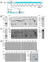
Fig. 1. Mapping the region of Pf332 that binds the RBC membrane skeleton
A. Schematic representation of full length Pf332 protein. The pf332 gene is comprised of two exons separated by an intron [30]. Residues 1 - 567 are encoded by the first exon of the pf332 gene and encodes a PEXEL motif (RSLAD), a Duffy Binding-Like domain (DBL; light shaded region) and a predicted transmembrane spanning domain (TM). Residues 568 - 6098 are encoded by the second exon of the pf332 gene and encode an extensive glutamic acid-rich region (darker shaded region). The relative locations of the twenty one fragments (F1 through F20, plus F18+19) that were expressed and purified as MBP fusion proteins and together represent the Pf332 protein sequence encoded by the second exon of pf332 are shown below the protein schematic. Relevant amino acid residue numbers are shown adjacent to the schematic. B. Purified MBP-Pf332 fusion proteins. 2μg (total protein) of each purified protein was resolved by SDS-PAGE and stained with Coomassie Brilliant Blue. The proteins are indicated by the protein fragment number above each lane. C. Each of the purified MBP-Pf332 fusion proteins (50ng total protein) was immunoblotted with anti-MBP antiserum to confirm the presence of the N-terminal MBP tag and demonstrate the apparent molecular mass of each protein when immunoblotted. D. IOV binding assay immunoblots. Samples stripped from IOV-coated wells (lanes headed by ‘+’) and BSA-coated wells (lanes headed by ‘−’) are indicated. The name of each Pf332 protein fragment analysed is shown above each pair of lanes. Pf332–F19 bound to IOVs, with only residual levels observed binding to BSA. The interaction positive control protein MBP-PfEMP3-F1a bound to IOVs and at much lower levels to BSA. MBP showed only residual binding to IOVs and did not bind to BSA. The immunoblotted assay samples, although separated into multiple panels here, were derived under identical and simultaneous experimental conditions.
Fig. 2
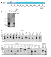
Fig. 2. RBC membrane skeleton association of Pf332
A. Schematic representation of full length Pf332 protein showing the relative locations of the twenty one Pf332 fragments investigated. B. Immunoblot detection of Pf332 in P. falciparum-IRBCs extracted with Triton X-100. Samples of IOVs and IOVs prepared from P. falciparum-IRBCs (pIOVs) were resolved alongside the TX100 insoluble fraction of P. falciparum 3D7 parasites in 0.5% (w/v) agarose/3.0% (w/v) acrylamide gels [36], before transferring to PVDF membrane and immunoblotting with anti-Pf332 antiserum. Pf332 was detected as a band of >584 kDa (indicated by the arrow) in the 3D7 TX100 insoluble fraction and in the pIOVs sample, but not in the IOVs prepared from normal human RBCs. Although originally described as a megadalton protein [25, 27], Pf332 is currently predicted to be a 700kDa protein, and was observed in our hands as having a molecular mass in excess of 584kDa. It is not uncommon for highly charged repetitive malaria proteins, such as Pf332, to resolve at anomalous higher-than-predicted molecular masses in SDS-PAGE [47]. C. TX100 extractions were preformed on lysed and resealed RBCs that had been incubated with 5 μM purified MBP-Pf332 fusion proteins, MBP alone, or with the binding control proteins MBP-PfEMP3 fusion proteins F1a or F5 (RBC membrane skeleton binding and non-binding regions, respectively; [15]). The TX100 soluble (S) and insoluble (I) fractions were resolved by SDS-PAGE and immunoblotted with anti-MBP antiserum. MBP-Pf332-F19 was detected in both the soluble and insoluble fractions. The region adjacent to –F19, –F18, was also detected in the insoluble fraction, but at lower levels than –F19. When -F18 and –F19 were combined into the same contiguous fusion protein (–F18+19), the protein was also detected in the insoluble fraction. As expected, the RBC membrane binding control protein MBP-PfEMP3-F1a was present in both the soluble and insoluble fractions, whereas the non-binding MBP-PfEMP3-F5 protein and MBP alone were present only in the soluble fractions. No significant immunoreactivity was observed in either fraction for TX100 extracted RBCs lysed and resealed in the absence of MBP or other MBP fusion proteins. The diversity of protein sizes and the number of bands per protein sample are reflective of the sizes of the Pf332 gene fragments expressed as MBP fusions, differences in their coding sequences, and the ability of E. coli to express these gene fragments as full length proteins.
Fig. 3
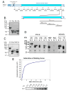
Fig. 3. Finer mapping of residues in Pf332-F19 that bind to the Membrane Skeleton
A. Schematic representation of full length Pf332 protein showing the relative locations on the twenty fragments investigated. To further define the binding residues contained within the 260 residue –F19 region, three smaller non-overlapping sub-fragments (-F19a, -F19b and –F19c) were expressed and purified as MBP fusion proteins. B. Purified MBP-Pf332 F19 sub-fragment fusion proteins. 2μg (total protein) of each purified protein was resolved by SDS-PAGE before staining with Coomassie Brilliant Blue. C. Each of the MBP-Pf332 F19 sub-fragment fusion proteins (50ng total protein) was immunoblotted with anti-MBP antiserum to confirm the presence of the N-terminal MBP tag and demonstrate the apparent molecular mass of each protein when immunoblotted. D. The TX100 soluble (S) and insoluble (I) fractions of lysed and resealed RBCs that had been incubated with 5 μM fusion protein were resolved by SDS-PAGE and immunoblotted with anti-MBP antiserum. MBP-Pf332-F19, and its sub-fragment proteins –F19a and –F19c were both detected in the Tx100 soluble and insoluble fractions, whereas the intervening sub-fragment –F19b was not. Neither MBP nor MBP-PfEMP3-F5 was detected in the insoluble fractions, whereas MBP-PfEMP3-F1a was. Together, these data map a membrane skeleton binding region of Pf332 to the 86 residues of –F19a. These data also suggest the presence of other residues within the downstream 72 residues of –F19c that may contribute to the overall protein interaction of Pf332 with the membrane skeleton. E. A saturation of binding curve was constructed from the densitometric data obtained from a representative IOV binding assay using serial dilutions of MBP-Pf332-F19 protein (titrated from 5.0 to 0.0 μM; panel below the graph). The half saturation concentration (Kd) was determined to be Kd = 0.40 μM.
Fig. 4
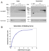
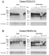
Fig. 4. Pf332-F19 binding to actin is saturable and specific
A. Actin co-sedimentation assays were performed using polymerised F-actin. Constant concentrations of F-actin (5 μM) were incubated with titrated serial dilutions of MBP-Pf332-F19 and centrifuged. The pellet and supernatant fractions were analysed by immunoblotting with anti-MBP and anti-actin antibodies and subsequent densitometry. MBP-Pf332-F19 pelleted with F-actin, whereas the non-binding MBP and MBP-Pf332-F7 proteins remained in the supernatant. B. A saturation of binding curve was constructed from the densitometric data obtained from a representative actin co-sedimentation assay using MBP-Pf332-F19 protein (Fig. 4A). The half saturation concentration (Kd) was determined to be Kd = 0.60 μM.
Fig. 5
Fig. 5. PfEMP3-F1a and Pf332-F19 bind to different regions of F-actin
Inhibition of binding to actin co-sedimentation assays were performed to determine if MBP-PfEMP3-F1a or MBP-Pf332-F19 bound to the same residues in actin. In these assays, F-actin (5 μM) was added to either MBP-PfEMP3-F1a (5 μM) or MBP-Pf332-F19 (5 μM) before the other protein (titrated from 5.0 to 0.0 μM) was added and the mixture subsequently incubated. Resultant pellet and supernatant fractions were immunoblotted using either anti-MBP antiserum or monoclonal anti-actin antibodies. A. Constant concentrations of F-actin (5 μM) and MBP-PfEMP3-F1a (5 μM; ~60 kDa) were incubated with titrated serial dilutions of MBP-Pf332-F19 (5.0 to 0.0 μM; ~85 kDa) and centrifuged. No inhibition of MBP-PfEMP3-F1a binding to F-actin in the presence of increasing concentrations of MBP-Pf332-F19 (0.0 to 5.0 μM) was detected in the pelleted fractions. Only residual levels of the negative interaction control proteins MBP (5.0 μM), MBP-Pf332-F7 (5.0 μM), MBP-Pf332-F5 (5.0 μM) pelleted with F-actin. The positive interaction controls MBP-PfEMP3-F1a (5.0 μM) and MBP-PF332-F19 (5.0 μM) each pelleting separately with F-actin were included. B. Constant concentrations of F-actin (5 μM) and MBP-Pf332-F19 (5 μM; ~85 kDa) were incubated with titrated serial dilutions of MBP-PfEMP3-F1a (5.0 to 0.0 μM; ~60 kDa) and centrifuged. No inhibition of MBP-Pf332-F19 binding to F-actin in the presence of increasing concentrations of MBP-PfEMP3-F1a (5.0 to 0.0 μM) was detected in the pelleted fractions. As above, only residual levels of the negative interaction control proteins MBP (5.0 μM), MBP-Pf332-F7 (5.0 μM), MBP-Pf332-F5 (5.0 μM) pelleted with F-actin. The positive interaction controls MBP-PfEMP3-F1a (5.0 μM) and MBP-PF332-F19 (5.0 μM) each pelleting separately with F-actin were included.

Similar articles

Cited by

References

    1. Snow RW, Guerra CA, Noor AM, Myint HY, Hay SI. The global distribution of clinical episodes of Plasmodium falciparum malaria. Nature. 2005;434:214–217. - PMC - PubMed
    1. Cooke BM, Mohandas N, Coppel RL. Malaria and the red blood cell membrane. Semin Hematol. 2004;41:173–188. - PubMed
    1. Walensky L, Narla M, Lux S., IV . Disorders of the Red Blood Cell Membrane. In: Handin R, Lux S IV, Stossel TP, editors. Blood: Princciples and Practice of Hematology. Lippincott, William and Wilkins; Philadelphia: 2003. pp. 1709–1858.
    1. Gascard P, Mohandas N. New insights into functions of erythroid proteins in nonerythroid cells. Curr Opin Hematol. 2000;7:123–129. - PubMed
    1. Karinch AM, Zimmer WE, Goodman SR. The Identification and Sequence of the Actin-binding Domain of Human Red Blood Cell β-Spectrin. J Biol Chem. 1990;265:11833–11840. - PubMed

Publication types

LinkOut - more resources