

Mice overexpressing corticotropin-releasing factor show brain atrophy and motor dysfunctions
- PMID: 20132869
- PMCID: PMC2848985
- DOI: 10.1016/j.neulet.2010.01.068
Mice overexpressing corticotropin-releasing factor show brain atrophy and motor dysfunctions
Abstract
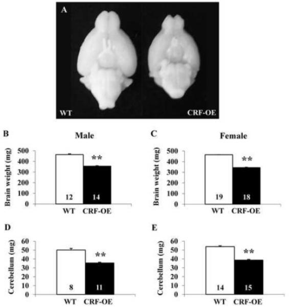
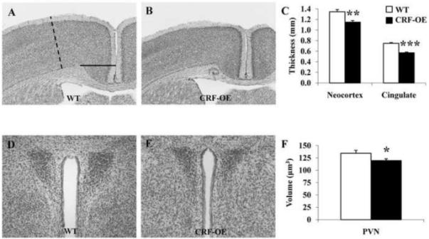
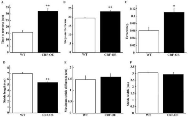
Chronic stress and persistently high glucocorticoid levels can induce brain atrophy. Corticotropin-releasing factor (CRF)-overexpressing (OE) mice are a genetic model of chronic stress with elevated brain CRF and plasma corticosterone levels and Cushing's syndrome. The brain structural alterations in the CRF-OE mice, however, are not well known. We found that adult male and female CRF-OE mice had significantly lower whole brain and cerebellum weights than their wild type (WT) littermates (347.7+/-3.6mg vs. 460.1+/-4.3mg and 36.3+/-0.8mg vs. 50.0+/-1.3mg, respectively) without sex-related difference. The epididymal/parametrial fat mass was significantly higher in CRF-OE mice. The brain weight was inversely correlated to epididymal/parametrial fat weight, but not to body weight. Computerized image analysis system in Nissl-stained brain sections of female mice showed that the anterior cingulate and sensorimotor cortexes of CRF-OE mice were significantly thinner, and the volumes of the hippocampus, hypothalamic paraventricular nucleus and amygdala were significantly reduced compared to WT, while the locus coeruleus showed a non-significant increase. Motor functions determined by beam crossing and gait analysis showed that CRF-OE mice took longer time and more steps to traverse a beam with more errors, and displayed reduced stride length compared to their WT littermates. These data show that CRF-OE mice display brain size reduction associated with alterations of motor coordination and an increase in visceral fat mass providing a novel animal model to study mechanisms involved in brain atrophy under conditions of sustained elevation of brain CRF and circulating glucocorticoid levels.
Copyright 2010 Elsevier Ireland Ltd. All rights reserved.
Figures
Similar articles
-
Corticotropin-releasing factor overexpression in mice abrogates sex differences in body weight, visceral fat, and food intake response to a fast and alters levels of feeding regulatory hormones.Biol Sex Differ. 2017 Jan 13;8:2. doi: 10.1186/s13293-016-0122-6. eCollection 2017. Biol Sex Differ. 2017. PMID: 28101317 Free PMC article.
-
Corticotropin releasing factor-overexpressing mouse is a model of chronic stress-induced muscle atrophy.PLoS One. 2020 Feb 12;15(2):e0229048. doi: 10.1371/journal.pone.0229048. eCollection 2020. PLoS One. 2020. PMID: 32049987 Free PMC article.
-
Corticotropin-releasing factor-overexpressing mice exhibit reduced neuronal activation in the arcuate nucleus and food intake in response to fasting.Endocrinology. 2009 Jan;150(1):153-60. doi: 10.1210/en.2008-0723. Epub 2008 Sep 11. Endocrinology. 2009. PMID: 18787020 Free PMC article.
-
Brain and Gut CRF Signaling: Biological Actions and Role in the Gastrointestinal Tract.Curr Mol Pharmacol. 2018;11(1):51-71. doi: 10.2174/1874467210666170224095741. Curr Mol Pharmacol. 2018. PMID: 28240194 Free PMC article. Review.
-
Corticotropin-releasing factor and the brain-gut motor response to stress.Can J Gastroenterol. 1999 Mar;13 Suppl A:18A-25A. doi: 10.1155/1999/375916. Can J Gastroenterol. 1999. PMID: 10202204 Review.
Cited by
-
Metabolic disturbances connecting obesity and depression.Front Neurosci. 2013 Oct 7;7:177. doi: 10.3389/fnins.2013.00177. Front Neurosci. 2013. PMID: 24109426 Free PMC article. Review.
-
Marital dissolution and cognition: The mediating effect of Aβ neuropathology.Alzheimers Dement (Amst). 2024 Oct 29;16(4):e70032. doi: 10.1002/dad2.70032. eCollection 2024 Oct-Dec. Alzheimers Dement (Amst). 2024. PMID: 39473984 Free PMC article.
-
Corticotropin-releasing factor receptor-1 antagonism mitigates beta amyloid pathology and cognitive and synaptic deficits in a mouse model of Alzheimer's disease.Alzheimers Dement. 2016 May;12(5):527-37. doi: 10.1016/j.jalz.2015.09.007. Epub 2015 Nov 7. Alzheimers Dement. 2016. PMID: 26555315 Free PMC article.
-
Increased tau phosphorylation and aggregation in the hippocampus of mice overexpressing corticotropin-releasing factor.J Alzheimers Dis. 2015;43(3):967-76. doi: 10.3233/JAD-141281. J Alzheimers Dis. 2015. PMID: 25125464 Free PMC article.
-
CRF receptor antagonist astressin-B reverses and prevents alopecia in CRF over-expressing mice.PLoS One. 2011 Feb 16;6(2):e16377. doi: 10.1371/journal.pone.0016377. PLoS One. 2011. PMID: 21359208 Free PMC article.
References
-
- McEwen BS. Possible mechanisms for atrophy of the human hippocampus. Mol. Psychiatry. 1997;2:255–262. - PubMed
-
- Galea LA, McEwen BS, Tanapat P, Deak T, Spencer RL, Dhabhar FS. Sex differences in dendritic atrophy of CA3 pyramidal neurons in response to chronic restraint stress. Neuroscience. 1997;81:689–697. - PubMed
-
- Sousa N, Cerqueira JJ, Almeida OF. Corticosteroid receptors and neuroplasticity. Brain Res. Rev. 2008;57:561–570. - PubMed
-
- Simmons NE, Do HM, Lipper MH, Laws ER., Jr. Cerebral atrophy in Cushing’s disease. Surg. Neurol. 2000;53:72–76. - PubMed
Publication types
MeSH terms
Substances
Grants and funding
LinkOut - more resources
Full Text Sources
Molecular Biology Databases
