

Mice overexpressing corticotropin-releasing factor show brain atrophy and motor dysfunctions
- PMID: 20132869
- PMCID: PMC2848985
- DOI: 10.1016/j.neulet.2010.01.068
Mice overexpressing corticotropin-releasing factor show brain atrophy and motor dysfunctions
Abstract
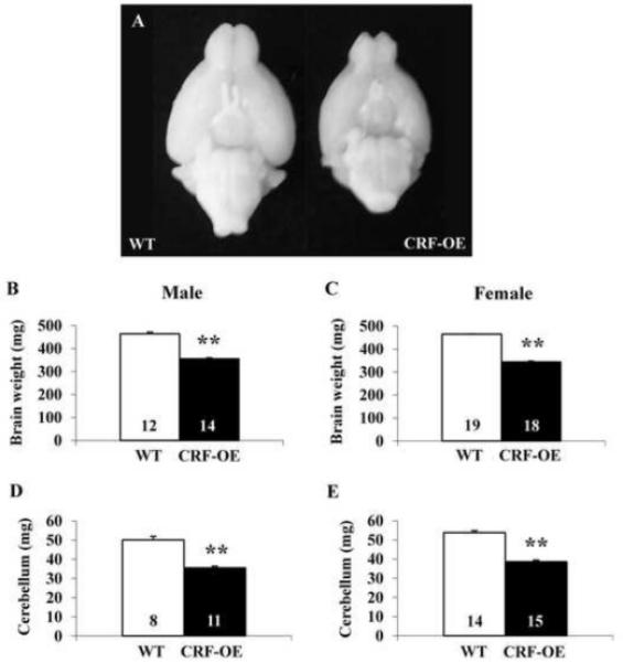
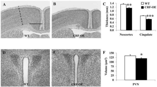
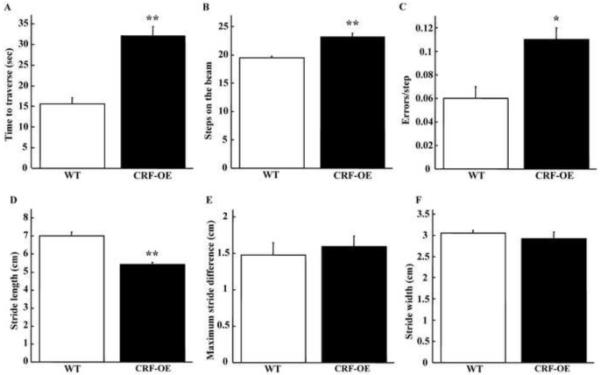
Chronic stress and persistently high glucocorticoid levels can induce brain atrophy. Corticotropin-releasing factor (CRF)-overexpressing (OE) mice are a genetic model of chronic stress with elevated brain CRF and plasma corticosterone levels and Cushing's syndrome. The brain structural alterations in the CRF-OE mice, however, are not well known. We found that adult male and female CRF-OE mice had significantly lower whole brain and cerebellum weights than their wild type (WT) littermates (347.7+/-3.6mg vs. 460.1+/-4.3mg and 36.3+/-0.8mg vs. 50.0+/-1.3mg, respectively) without sex-related difference. The epididymal/parametrial fat mass was significantly higher in CRF-OE mice. The brain weight was inversely correlated to epididymal/parametrial fat weight, but not to body weight. Computerized image analysis system in Nissl-stained brain sections of female mice showed that the anterior cingulate and sensorimotor cortexes of CRF-OE mice were significantly thinner, and the volumes of the hippocampus, hypothalamic paraventricular nucleus and amygdala were significantly reduced compared to WT, while the locus coeruleus showed a non-significant increase. Motor functions determined by beam crossing and gait analysis showed that CRF-OE mice took longer time and more steps to traverse a beam with more errors, and displayed reduced stride length compared to their WT littermates. These data show that CRF-OE mice display brain size reduction associated with alterations of motor coordination and an increase in visceral fat mass providing a novel animal model to study mechanisms involved in brain atrophy under conditions of sustained elevation of brain CRF and circulating glucocorticoid levels.
Copyright 2010 Elsevier Ireland Ltd. All rights reserved.
Figures
References
-
- McEwen BS. Possible mechanisms for atrophy of the human hippocampus. Mol. Psychiatry. 1997;2:255–262. - PubMed
-
- Galea LA, McEwen BS, Tanapat P, Deak T, Spencer RL, Dhabhar FS. Sex differences in dendritic atrophy of CA3 pyramidal neurons in response to chronic restraint stress. Neuroscience. 1997;81:689–697. - PubMed
-
- Sousa N, Cerqueira JJ, Almeida OF. Corticosteroid receptors and neuroplasticity. Brain Res. Rev. 2008;57:561–570. - PubMed
-
- Simmons NE, Do HM, Lipper MH, Laws ER., Jr. Cerebral atrophy in Cushing’s disease. Surg. Neurol. 2000;53:72–76. - PubMed
Publication types
MeSH terms
Substances
Grants and funding
LinkOut - more resources
Full Text Sources
Molecular Biology Databases
