Comparative Study
. 2011 Dec;32(12):2287-98.
doi: 10.1016/j.neurobiolaging.2010.01.003. Epub 2010 Feb 4.

Association of AICD and Fe65 with Hirano bodies reduces transcriptional activation and initiation of apoptosis

Affiliations
Comparative Study

Association of AICD and Fe65 with Hirano bodies reduces transcriptional activation and initiation of apoptosis

Sangdeuk Ha et al. Neurobiol Aging. 2011 Dec.

Abstract

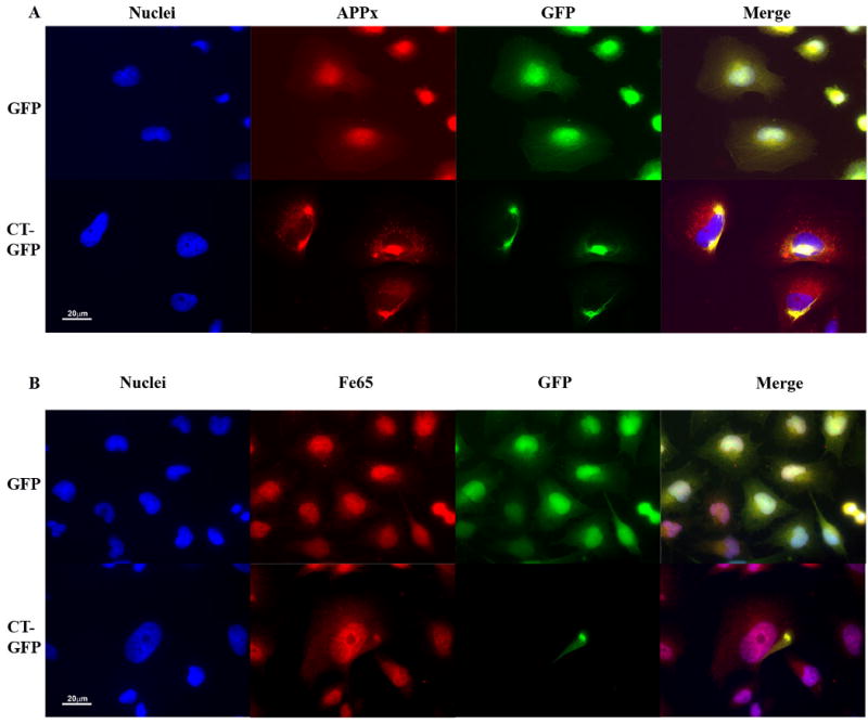
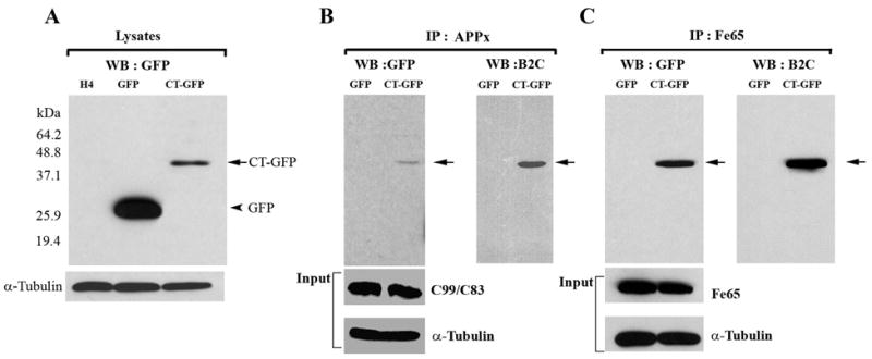
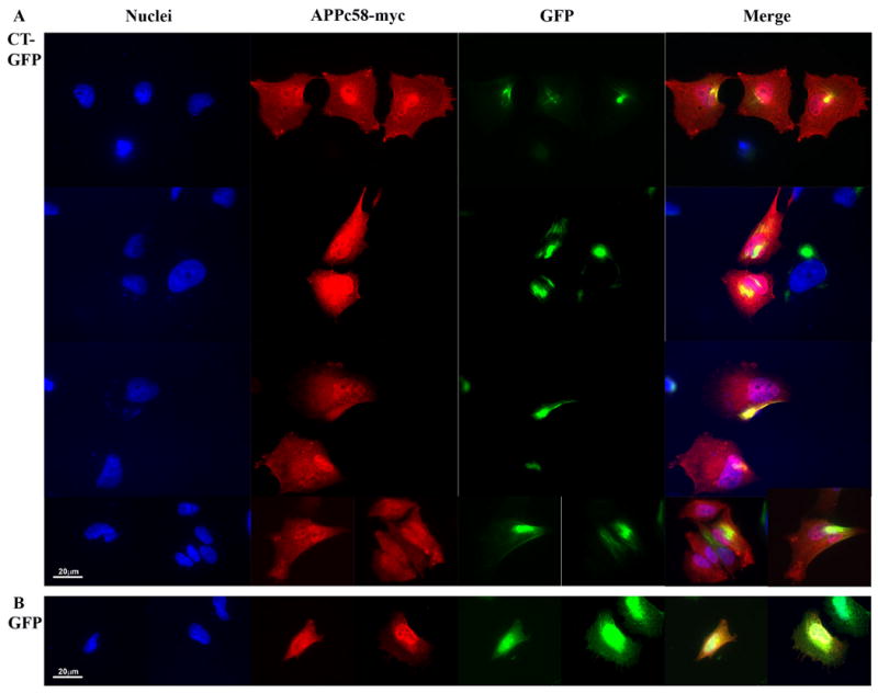
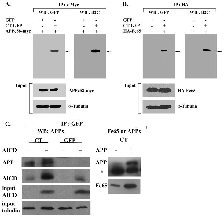
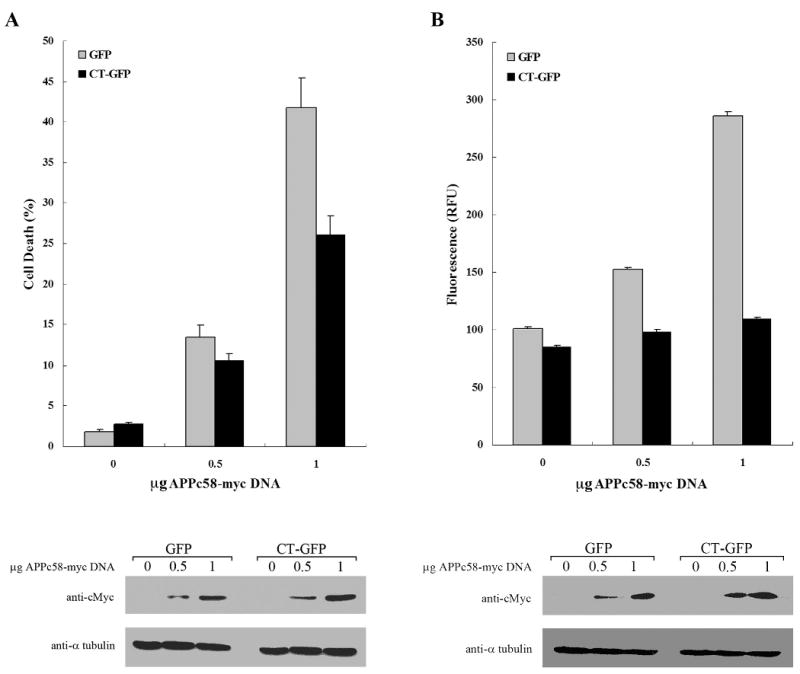
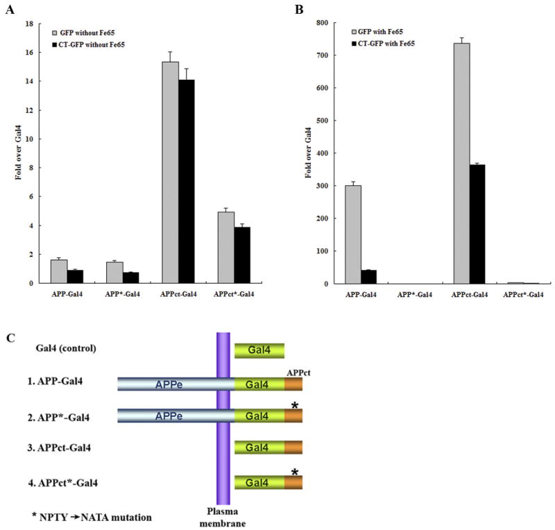
Hirano bodies are cytoplasmic inclusions predominantly found in the central nervous system associated with various conditions including aging and Alzheimer's disease (AD). Since most studies of Hirano bodies have been performed in post-mortem samples, the physiological roles of Hirano bodies have not been investigated. Astrocytoma H4 cells were employed to test the hypothesis that Hirano bodies interact with and modulate signaling by the C-terminal fragment of amyloid-β precursor protein (AICD). We demonstrated by immunofluorescence and immunoprecipitation that model Hirano bodies accumulate AICD. Since stimulation of transcription by AICD is dependent on its interaction with the nuclear adaptor protein Fe65, we examined localization of Fe65, and employed a dual luciferase reporter assay to test the effects of Hirano bodies on AICD- and Fe65-dependent modulation of gene expression. We find that both AICD and Fe65 are co-localized in model Hirano bodies. Model Hirano bodies also down-regulate both AICD-dependent apoptosis and AICD- and Fe65-dependent transcriptional activity. Thus, association of AICD and Fe65 with Hirano bodies impedes their function in promoting apoptosis and modulating transcription.


Conflict of interest statement

Disclosure Statement: there are no conflicts of interest

Figures

Figure 1
Figure 1
The C-terminal domain of the amyloid-β precursor protein is co-localized with Hirano bodies. H4 cells stably expressing GFP or CT-GFP were stained for the presence of endogenous forms of APP that include the C-terminus (APPx) (A) or Fe65 (B), and visualized by TRITC-conjugated secondary antibodies (red). DNA was stained with Hoechst dye (blue). A. APPx strongly localized with the nucleus and perinuclear region with limited diffuse cytoplasmic staining in GFP stable cells, and showed extensive co-localization with Hirano bodies in the cytoplasm in CT-GFP stable cells. Areas of co-localization appear yellow in the merged image. B. Fe65 co-localized extensively with nuclei in GFP stable cells and is partitioned between the nucleus and Hirano bodies in CT-GFP stable cells. Scale bar = 20 μm.
Figure 2
Figure 2
Hirano bodies bind to C-terminal fragments of APP (APP695, APPC99/APPC83 and APPC57-59) and Fe65. A. Cell lysates from wild type, GFP, and CT-GFP stable H4 cells were analyzed by western blot using antibody against GFP. To verify equal loading of the different cell lysates, α-tubulin was employed as a control. The 46 kDa CT-GFP fusion protein (arrow) and 27 kDa GFP protein (arrowhead) are detected as single bands. Marker proteins are 64.2, 48.8, 37.1, 25.9, and 19.4 kDa. B, C. Cell lysates obtained from GFP and CT-GFP stable H4 cells were immunoprecipitated with an antibody against the C-terminal portion of APP (anti-APPx) or with anti-Fe65 antibody, respectively. Products were detected with either anti-GFP or B2C, a monoclonal antibody against 34 kDa protein. To verify equal loading of the different cell lysates, α-tubulin was employed as a control as well as APPx (panel B) and Fe65 (panel C). The immunoprecipitate fraction in panel B contained full length APP (not shown) as well as C99/C83. The results show a specific interaction of APPx (B) and Fe65 (C) with CT-GFP in model Hirano bodies, but not with GFP.
Figure 3
Figure 3
The C-terminal domain of the amyloid-β precursor protein is co-localized with Hirano bodies. H4 stable cells expressing (A) CT-GFP or (B) GFP were transiently transfected with APPc58-myc and visualized by anti-myc, and TRITC-conjugated secondary antibody (red) for APPc58-myc. DNA was stained with Hoechst dye (blue). A. Areas of specific co-localization of APPc58-myc with Hirano bodies in the cytoplasm in CT-GFP stable cells appear yellow in the merged images. B. APPc58-myc is strongly localized in the nucleus in GFP stable cells. Scale bar = 20 μm.
Figure 4
Figure 4
Co-localization of AICD and Fe65 with Hirano bodies. Lysates from cells transfected with APPc58-myc (A, C) or HA-Fe65 (B) or APP695 (C) were immunoprecipitated with anti-myc antibody (A) or anti-HA antibody (B) or anti-GFP (C). The immunoprecipitated proteins were analyzed by western blot with either anti-GFP or anti-34 kDa (B2C) antibody or anti-APPct or anti-Fe65 (B). To verify equal loading of the different cell lysates, α-tubulin was employed as a control as well as APPc58-myc in A, C and HA-Fe65 in B. The results show a specific interaction of APPc58 (A,C), Fe65 (B,C), and APP695 with CT-GFP in model Hirano bodies, but not with GFP in control cells. The * in C indicates the heavy chain of IgG arising from the GFP antibody used for the immunoprecipitation.
Figure 5
Figure 5
Hirano bodies significantly decrease AICD-induced apoptosis in H4 cells. H4 stable cells stably expressing GFP or CT-GFP were transiently transfected with APPc58-myc DNA (0, 0.5, or 1 μg DNA), and examined at 24 hours for cell viability (A) and caspase 3/7 activation (B). A. Cells were stained with propidium iodide (PI) and Hoechst dye 33342 to assess cell viability. CT-GFP stable cells with Hirano bodies showed a lower percentage of cell death than GFP stable cells (p < 0.001). B. Caspase-3 activity was measured using a fluorescence assay that was normalized to β-galactosidase activity as a control for transfection efficiency of APPc58-myc. Caspase-3 activity represented by relative fluorescence units (RFU) was significantly decreased in CT-GFP stable cells compared to GFP stable cells (p < 0.001). Data shown are mean ± S.E.M. of triplicate measurements, and repeated in three independent experiments. Cell lysates transiently transfected with various concentrations of APPc58-myc DNA were examined by immunoblotting with anti-c-Myc antibodies (bottom panel) to verify that expression of APPc58-myc increased with the amount of DNA transfected.
Figure 6
Figure 6
Fe65-dependent transcriptional activity of APP-Gal4 and APPct-Gal4 is down regulated in HEK293T stable cells with Hirano bodies. HEK293T stable cells (1×10⁴ cells) expressing GFP or CT-GFP were co-transfected with 0.1 μg of pG5E1B-luc, 0.02 μg of pRL-TK and one of the following 5 different Gal4 constructs (C): 0.1 μg of pMst (Gal4), 0.1 μg of pMst-APP695 (APP-Gal4), 0.1 μg of pMst-APP695* (APP*-Gal4), 0.1 μg of pMst-APPct (APPct-Gal4), and 0.1 μg of pMst-APPct* (APPct*-Gal4) in the absence (A) or presence (B) of 0.1 μg of HA-Fe65. Plasmids with * contain the NATA mutation instead of NPTY in the C-terminal domain of APP that binds to Fe65. Transactivation activity was normalized to the level of transactivation of control Gal4. APP-Gal4 and APPct-Gal4 induced reporter expression was greatly enhanced by the presence of Fe65 (note the different scale bars in panels A and B). In the presence of Fe65, reporter expression was significantly lower in CT-GFP cells as compared to GFP cells (p < 0.001), and was significantly greater for APP-Gal4 and APPct-Gal4 than for the corresponding constructs with the NATA mutation (marked with *) in the Fe65 binding motif. Data shown are mean ± S.E.M. of triplicate measurements and repeated in three independent experiments.

Similar articles

Cited by

References

    1. Alves da Costa C, Sunyach C, Pardossi-Piquard R, Sevalle J, Vincent B, Boyer N, Kawarai T, Girardot N, St George-Hyslop P, Checler F. Presenilin-dependent gamma-secretase-mediated control of p53-associated cell death in Alzheimer's disease. J Neurosci. 2006;26:6377. - PMC - PubMed
    1. Ballatore C, Lee VM, Trojanowski JQ. Tau-mediated neurodegeneration in Alzheimer's disease and related disorders. Nat Rev Neurosci. 2007;8:663–672. - PubMed
    1. Banwait S, Galvan V, Zhang J, Gorostiza OF, Ataie M, Huang W, Crippen D, Koo EH, Bredesen DE. C-terminal cleavage of the amyloid-beta protein precursor at Asp664: a switch associated with Alzheimer's disease. J Alzheimer's Disease. 2008;13:1–16. - PMC - PubMed
    1. Butterfield DA, Griffin S, Munch G, Pasinetti GM. Amyloid beta-peptide and amyloid pathology are central to the oxidative stress and inflammatory cascades under which Alzheimer's disease brain exists. J Alzheimer's Dis. 2002;4:193. - PubMed
    1. Cao X, Südhof TC. A transcriptionally active complex of APP with Fe65 and histone acetyltransferase Tip60. Science. 2001;293:115. - PubMed

Publication types

MeSH terms

Substances