Review
. 2010 Apr;20(4):187-95.
doi: 10.1016/j.tcb.2010.01.001. Epub 2010 Feb 3.

ADF/cofilin: a functional node in cell biology

Affiliations
Review

ADF/cofilin: a functional node in cell biology

Barbara W Bernstein et al. Trends Cell Biol. 2010 Apr.

Abstract

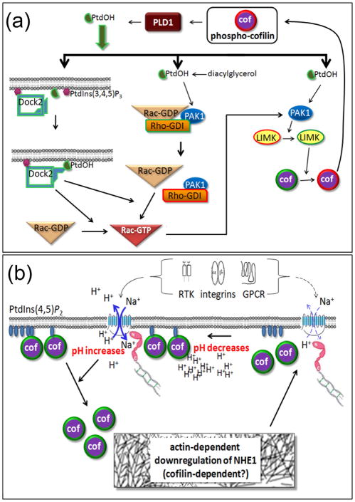
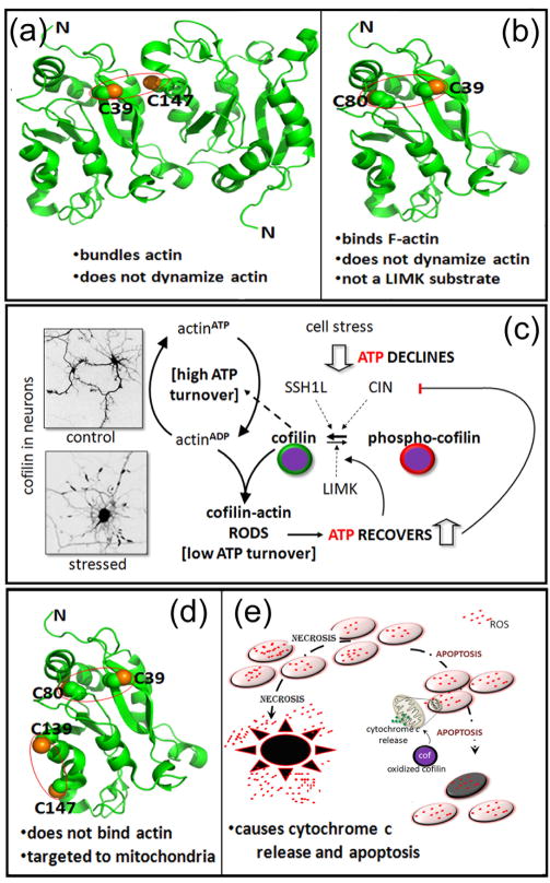
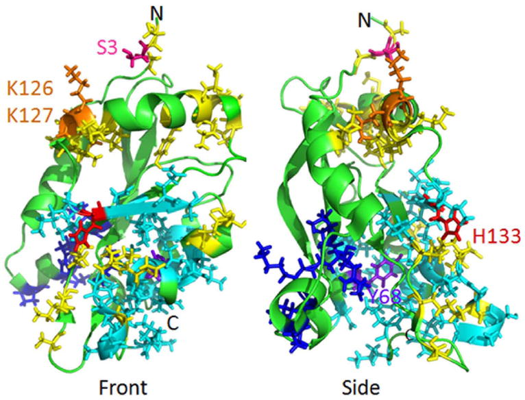
Recent findings have significantly expanded our understanding of the regulation of actin-depolymerizing factor (ADF)/cofilin proteins and the profound multifaceted impact that these well-established regulators of actin dynamics have on cell biology. In this review we discuss new aspects of previously documented regulation, such as phosphorylation, but also cover novel recently established modes of regulation and functions of ADF (also known as destrin)/cofilin. We now understand that their activity responds to a vast array of inputs far greater than previously appreciated and that these proteins not only feed back to the crucially important dynamics of actin, but also to apoptosis cascades, phospholipid metabolism, and gene expression. We argue that this ability to respond to physiological changes by modulating those same changes makes the ADF/cofilin protein family a homeostatic regulator or 'functional node' in cell biology.


Figures

Figure 1
Figure 1. Diverse functions of cofilin including actin dynamics and beyond
(a) Diagram showing several of the functions of cofilin (cof) within the cell that are described in more detail throughout the text. (b) Many of the factors that regulate cofilin activity are themselves modulated by cofilin and phospho-cofilin. Thus cofilin has a homeostatic role. (c) An example of homeostatic feedback regulation is the oxidative stress cycle. Reactive oxygen species (ROS), made as a result of signaling pathways or from other sources, activate cofilin phosphatases through different mechanisms discussed in the text. The resulting active cofilin can restructure the cortical actin and receptor interactions, modulating signaling. As with all figures in this article, symbols surrounded by a red line are inactive and those surrounded by a green line are active.
Figure 2
Figure 2. Recently discovered roles for cofilin in lipid metabolism and signaling and in pH-regulated actin remodeling
(a) Phospho-cofilin is an activator of phospholipase D1 (PLD1) and thus a regulator of phosphatidic acid (PtdOH) production. PtdOH plays multiple roles in the activation of Rac1, a Rho family GTPase, as well as directly activating PAK1, a kinase that is also downstream of active Rac1. PAK1 is an activator of the cofilin kinase LIMK1. Thus a phospho-cofilin/PtdOH positive feedback cycle exists. (b) Many receptors along with intracellular acidity activate the Na+/H+ exchanger (NHE1), linked via molecules in the ezrin/moesin/radixin family (shown in pink) to the actin cytoskeleton. The influx of Na+ and efflux of H+ locally elevates pH that enhances the release of cofilin from its inhibitory membrane binding to PtdIns(3,4)P2. Released cofilin may then contribute to actin remodeling that can decrease the flux of the NHE1 and lower pH. Thus cofilin is a likely regulator of pH homeostasis.
Figure 3
Figure 3. Cofilin as a mediator of oxidative stress
(a) Hypothetical model of a cofilin dimer linked by a disulfide bridge between C39 of one cofilin and C147 of another. This dimer promotes actin bundling in vitro, has been formed by oxidized glutathione [49] but was not detected in vivo following cell exposure to taurine chloramine, the major oxidant of neutrophils [14]. Instead, taurine chloramine induces one or two intramolecular disulfide bonds from the four cofilin cysteine residues. (b) The intramolecular disulfide bond between C39 and C80. The two residues are not close enough to form a disulfide bond without some alteration in the protein structure, which causes a loss of actin dynamizing activity without eliminating F-actin binding. (c) Cofilin-actin rods form when abnormally high levels of active cofilin and ADP-actin are reached, as in response to oxidative stress and ATP decline. The inverted images show cofilin immunostained neurons under control and stressed conditions. Rods can sequester a large percentage of the active cofilin, thus slowing the decline in ATP brought about by cofilin-stimulated actin turnover and its associated ATP hydrolysis. That turnover consumes considerable ATP. The ability to maintain some critical level of ATP is necessary for the cell to recover as stress subsides. Thus cofilin contributes to energy homeostasis. (d) The fully oxidized cofilin, with disulfide bonds between C39–C80 and C139–C147, does not bind to F-actin but is targeted to mitochondrial outer membrane. Preliminary modeling of the structure showed a disruption in the alpha-5 helix, which includes C147. Two F-actin-binding residues (Box 3) reside nearby, and the alteration in structure probably accounts for the loss of F-actin binding. (e) Fully oxidized cofilin (d) is targeted to the mitochondrial outer membrane where it causes leakage of cytochrome c and activation of an apoptotic cascade. Apoptosis causes much less tissue damage than necrosis, a process responsible for inflammation, increased ROS, and extensive cell death.
Figure I
Figure I

Similar articles

Cited by

References

    1. Hotulainen P, et al. Actin-depolymerizing factor and cofilin-1 play overlapping roles in promoting rapid F-actin depolymerization in mammalian nonmuscle cells. Mol Biol Cell. 2005;16:649–664. - PMC - PubMed
    1. Gurniak CB, et al. The actin depolymerizing factor n-cofilin is essential for neural tube morphogenesis and neural crest cell migration. Dev Biol. 2005;278:231–241. - PubMed
    1. Ikeda S, et al. Aberrant actin cytoskeleton leads to accelerated proliferation of corneal epithelial cells in mice deficient for destrin (actin depolymerizing factor) Hum Mol Genet. 2003;12:1029–1036. - PubMed
    1. Van Troys M, et al. Ins and outs of ADF/cofilin activity and regulation. Eur J Cell Biol. 2008;87:649–667. - PubMed
    1. Andrianantoandro E, Pollard TD. Mechanism of actin filament turnover by severing and nucleation at different concentrations of ADF/cofilin. Mol Cell. 2006;24:13–23. - PubMed

Publication types