

Review
doi: 10.3174/ajnr.A2002.
Epub 2010 Feb 4.
MR neurography of neuromas related to nerve injury and entrapment with surgical correlation
Affiliations
- PMID: 20133388
- PMCID: PMC7966120
- DOI: 10.3174/ajnr.A2002
Item in Clipboard
Review
MR neurography of neuromas related to nerve injury and entrapment with surgical correlation
AJNR Am J Neuroradiol.
2010 Sep.
Abstract
MR imaging of peripheral nerves has been described in relation to abnormalities such as nerve injury, entrapment, and neoplasm. Neuroma formation is a known response to peripheral nerve injury, and here we correlate the MRN appearance of postinjury neuroma formation with intraoperative findings. We also present the MR imaging features of surgical treatment with a synthetic nerve tube and nerve wrap on postoperative follow-up imaging.
Figures
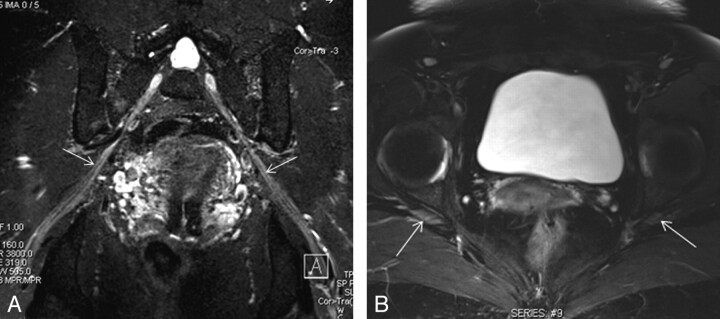
3D STIR SPACE in an oblique coronal reconstruction (A) and axial T2 SPAIR (B) images demonstrate normal fascicular appearance of the sciatic nerves (arrows).
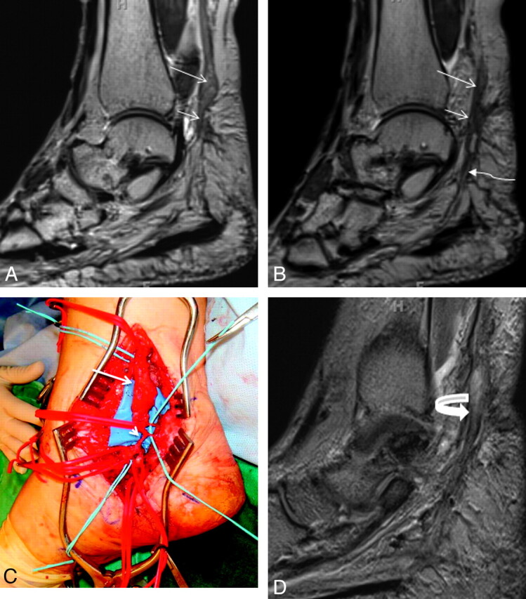
A 46-year-old woman with a history of attempted right tarsal tunnel release presented with persistent foot and ankle pain with numbness in the medial plantar distribution. A and B, Sequential sagittal T2 SPACE images demonstrate a spindle-shaped NIC involving the distal tibial nerve (long arrows). Notice the attenuated appearance of the medial (short arrows) and lateral (wavy arrow) plantar nerves. C, Intraoperative photo confirmed the NIC (long arrow) and small proximal medial and lateral plantar nerves entrapped in scarring (short arrow). D, Extensive neurolysis was performed and a nerve wrap was placed. Follow-up sagittal T2 SPACE MR image shows the wrap as a hypointense covering around the tibial and proximal medial plantar nerve (curved arrow), with residual hyperintensity of the nerves.
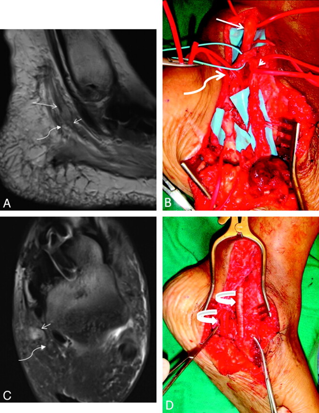
A 48-year-old woman with a history of attempted left tarsal tunnel release and midfoot fusion presented with severe pain in the bottom of the foot with toe flexion weakness. A and C, Sagittal T2 SPACE (A) and axial T2 SPAIR images (C) demonstrate a spindle-shaped NIC involving the tibial nerve (arrow), medial plantar nerve (short arrow), and relatively less involved lateral plantar nerve (wavy arrow) entrapped in surrounding scarring. Extensive denervation edema and atrophy of plantar muscles were also identified (not shown). B, The findings correlate well with intraoperative photography. D, Collagen-based nerve wraps are placed around the neurolysed segments of medial and lateral plantar nerves (curved arrows), with successful recovery.
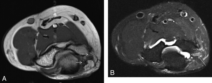
A 42-old-woman with tingling and numbness in the ulnar side of the right hand. Axial T1 (A) and T2 SPAIR (B) images show a hyperintense ulnar nerve (long arrows) entrapped at the cubital tunnel due to focal fibrosis (short arrows) related to previous injury. No denervation atrophy was seen at the time of imaging, and clinical as well as electromyography findings were in keeping with neurapraxia.
Benign peripheral nerve sheath tumor of the sciatic nerve shows the typical split fat sign (arrow) on coronal T1 (A), target sign (short arrow) on coronal STIR (B), and nodular enhancement (curved arrow) on a postcontrast coronal T1 fat-saturated image (C).
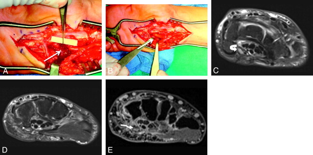
A 49-year-old woman with a history of previous carpal tunnel surgery and median nerve injury. A, The median nerve was re-explored in the hand followed by neurolysis and nerve tube placement (short arrows). The patient did not recover nerve function following surgery. B and C, MR imaging examination 5 months after surgery shows no significant nerve regeneration and empty fluid-filled nerve tubes (curved arrows). D, Re-exploration demonstrated end-bulb neuromas (wavy arrow), and nerve grafting was performed.
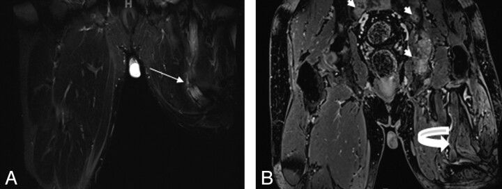
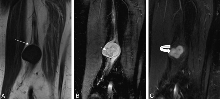
A 50-year-old man with previous left leg amputation for epitheloid sarcoma presents with multiple bony metastases. A and B, Notice an enlarged nonenhancing hyperintense sciatic nerve with an amputation end-bulb neuroma on coronal STIR (arrow, A) and postcontrast T1 3D gradient recalled-echo (curved arrow, B) images.
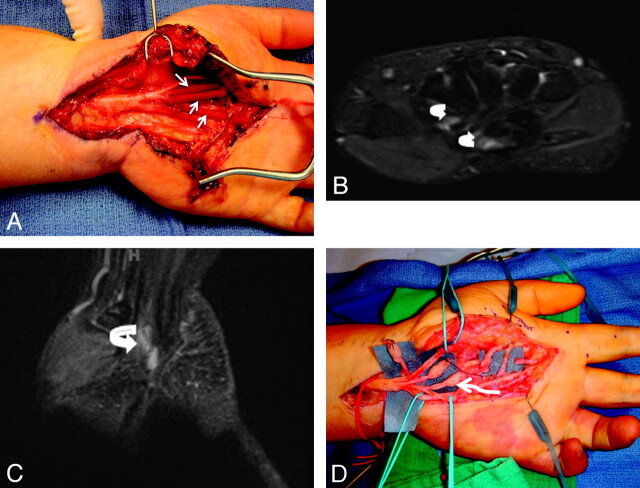
A 48-year-old woman presented with a claw hand following injury to the ulnar nerve during ganglion cyst removal from Guyon canal. A and B, During repeat surgery, the severed nerve is sutured to the ends of a neurotube (arrows). C−E, MRN examination was performed 1 month after the surgery. Axial T2 SPAIR image (C) shows an enlarged and hyperintense ulnar nerve proximally related to long-standing obstruction of axoplasmic flow (curved arrow) and postoperative changes. At the level of the hook of the hamate, minimal filling of the neural tube with nonenhancing tissue (arrowhead on the axial T2 SPAIR image in D, and wavy arrow in the postcontrast 3D T1 fat-saturated image in E) is hypothesized to represent early nerve sprouts.
References
-
- Bencardino JT, Rosenbert ZS. Entrapment neuropathies of the upper extremity. In: Stoller DW.ed. Magnetic Resonance Imaging in Orthopaedics and Sports Medicine. Baltimore: Lippincott Williams & Wilkins; 2006
-
- Chen ZL, Yu WM, Strickland S. Peripheral regeneration. Annu Rev Neurosci 2007;30:209–33 - PubMed
-
- Aagaard BD, Maravilla KR, Kliot M. Magnetic resonance neurography: magnetic resonance imaging of peripheral nerves. Neuroimaging Clin N Am 2001;11:131–46 - PubMed
-
- Ide C. Peripheral nerve regeneration. Neurosci Res 1996;25:101–21 - PubMed
Publication types
MeSH terms
LinkOut - more resources
Full Text Sources
Medical