

Cerebral white matter recovery in abstinent alcoholics--a multimodality magnetic resonance study
- PMID: 20133395
- PMCID: PMC2850577
- DOI: 10.1093/brain/awp343
Cerebral white matter recovery in abstinent alcoholics--a multimodality magnetic resonance study
Abstract
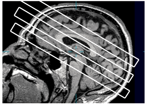
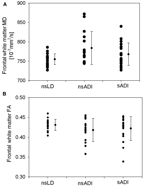
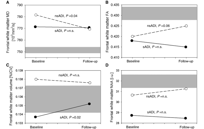
Most previous neuroimaging studies of alcohol-induced brain injury and recovery thereof during abstinence from alcohol used a single imaging modality. They have demonstrated widespread microstructural, macrostructural or metabolite abnormalities that were partially reversible with abstinence, with the cigarette smoking potentially modulating these processes. The goals of this study were to evaluate white matter injury and recovery thereof, simultaneously with diffusion tensor imaging, magnetic resonance imaging and spectroscopy in the same cohort; and to evaluate the relationships between outcome measures of similar regions. We scanned 16 non-smoking and 20 smoking alcohol-dependent individuals at 1 week of abstinence from alcohol and 22 non-smoking light drinkers using a 1.5 T magnetic resonance scanner. Ten non-smoking alcohol-dependent individuals and 11 smoking alcohol-dependent individuals were re-scanned at 1 month of abstinence. All regional diffusion tensor imaging, magnetic resonance imaging and spectroscopic outcome measures were calculated over comparable volumes of frontal, temporal, parietal and occipital white matter. At 1 week of abstinence and relative to non-smoking light drinkers, non-smoking alcohol-dependent individuals had higher mean diffusivity in frontal, temporal and parietal white matter (all P<0.008), whereas smoking alcohol-dependent individuals had elevated mean diffusivity only in frontal white matter (P=0.03). Smoking alcohol-dependent individuals demonstrated lower concentrations of N-acetyl-aspartate (a marker of neuronal viability) in frontal white matter (P=0.03), whereas non-smoking alcohol-dependent individuals had lower N-acetyl-aspartate in parietal white matter (P=0.05). These abnormalities were not accompanied by detectable white matter atrophy. However, the patterns of white matter recovery were different between non-smoking alcohol-dependent individuals and smoking alcohol-dependent individuals. In non-smoking alcohol-dependent individuals, the increase in fractional anisotropy of temporal white matter (P=0.003) was accompanied by a pattern of decreases mean diffusivity in all regions over 1 month of abstinence; no corresponding changes were observed in smoking alcohol-dependent individuals. In contrast, a pattern of white matter volume increase in frontal and temporal lobes was apparent in smoking alcohol-dependent individuals but not in non-smoking alcohol-dependent individuals. These results were not accompanied by significant changes in metabolite concentrations. Finally, there were no consistent patterns of association between measures obtained with different imaging modalities, either cross-sectionally or longitudinally. These data demonstrate significant white matter improvements with abstinence from alcohol, reflected either as microstructural recovery or volumetric increases that depend on the smoking status of the participants. We believe our results to be important, as they demonstrate that use of a single imaging modality provides an incomplete picture of neurobiological processes associated with alcohol-induced brain injury and recovery thereof that may even lead to improper interpretation of results.
Figures
Similar articles
-
Callosal white matter microstructural recovery in abstinent alcoholics: a longitudinal diffusion tensor imaging study.Alcohol Clin Exp Res. 2012 Nov;36(11):1922-31. doi: 10.1111/j.1530-0277.2012.01808.x. Epub 2012 May 2. Alcohol Clin Exp Res. 2012. PMID: 22551067 Free PMC article.
-
The impact of chronic cigarette smoking on recovery from cortical gray matter perfusion deficits in alcohol dependence: longitudinal arterial spin labeling MRI.Alcohol Clin Exp Res. 2009 Aug;33(8):1314-21. doi: 10.1111/j.1530-0277.2009.00960.x. Epub 2009 Apr 30. Alcohol Clin Exp Res. 2009. PMID: 19413652 Free PMC article.
-
Effects of abstinence on the brain: quantitative magnetic resonance imaging and magnetic resonance spectroscopic imaging in chronic alcohol abuse.Alcohol Clin Exp Res. 2001 Nov;25(11):1673-82. Alcohol Clin Exp Res. 2001. PMID: 11707642
-
Magnetic resonance imaging of the living brain: evidence for brain degeneration among alcoholics and recovery with abstinence.Alcohol Res Health. 2008;31(4):362-76. Alcohol Res Health. 2008. PMID: 23584010 Free PMC article. Review.
-
Neuroimaging in cerebrovascular disorders: measurement of cerebral physiology after stroke and assessment of stroke recovery.Semin Nucl Med. 2003 Jan;33(1):56-76. doi: 10.1053/snuc.2003.127293. Semin Nucl Med. 2003. PMID: 12605357 Review.
Cited by
-
Neuropsychological dysfunctions among chronic schizophrenia patients, alcohol dependence cases, and normal subjects: A comparative study.Ind Psychiatry J. 2020 Jan-Jun;29(1):105-122. doi: 10.4103/ipj.ipj_70_20. Epub 2020 Nov 7. Ind Psychiatry J. 2020. PMID: 33776284 Free PMC article.
-
Therapeutic Effects of Myriocin in Experimental Alcohol-Related Neurobehavioral Dysfunction and Frontal Lobe White Matter Biochemical Pathology.J Behav Brain Sci. 2022 Feb;12(2):23-42. doi: 10.4236/jbbs.2022.122003. Epub 2022 Feb 10. J Behav Brain Sci. 2022. PMID: 36815096 Free PMC article.
-
Caudate gray matter volumes and risk of relapse in Type A alcohol-dependent patients: A 7-year MRI follow-up study.Front Psychiatry. 2023 Feb 15;14:1067326. doi: 10.3389/fpsyt.2023.1067326. eCollection 2023. Front Psychiatry. 2023. PMID: 36873223 Free PMC article.
-
Harnessing neuroplasticity for clinical applications.Brain. 2011 Jun;134(Pt 6):1591-609. doi: 10.1093/brain/awr039. Epub 2011 Apr 10. Brain. 2011. PMID: 21482550 Free PMC article.
-
Bilateral fronto-parietal integrity in young chronic cigarette smokers: a diffusion tensor imaging study.PLoS One. 2011;6(11):e26460. doi: 10.1371/journal.pone.0026460. Epub 2011 Nov 1. PLoS One. 2011. PMID: 22069452 Free PMC article.
References
-
- Agartz I, Brag S, Franck J, Hammarberg A, Okugawa G, Svinhufvud K, et al. MR volumetry during acute alcohol withdrawal and abstinence: a descriptive study. Alcohol Alcohol. 2003;38:71–8. - PubMed
-
- Alexander AL, Hasan KM, Lazar M, Tsuruda JS, Parker DL. Analysis of partial volume effects in diffusion-tensor MRI. Magn Reson Med. 2001;45:770–80. - PubMed
-
- Alonso JR, Cardellach F, Casademont J, Miro O. Reversible inhibition of mitochondrial complex IV activity in PBMC following acute smoking. Eur Respir J. 2004;23:214–18. - PubMed
-
- Bartsch AJ, Homola G, Biller A, Smith SM, Weijers HG, Wiesbeck GA, et al. Manifestations of early brain recovery associated with abstinence from alcoholism. Brain. 2007;130:36–47. - PubMed
-
- Bartzokis G, Beckson M, Lu PH, Nuechterlein KH, Edwards N, Mintz J. Age-related changes in frontal and temporal lobe volumes in men: a magnetic resonance imaging study. Arch Gen Psychiatry. 2001;58:461–5. - PubMed
