. 2010 May;108(5):1336-46.
doi: 10.1152/japplphysiol.00477.2009. Epub 2010 Feb 4.

Serotonergic projection from nucleus raphe pallidus to rostral ventrolateral medulla modulates cardiovascular reflex responses during acupuncture

Affiliations

Serotonergic projection from nucleus raphe pallidus to rostral ventrolateral medulla modulates cardiovascular reflex responses during acupuncture

Ali Moazzami et al. J Appl Physiol (1985). 2010 May.

Abstract

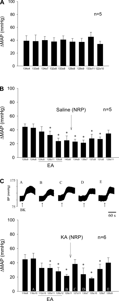
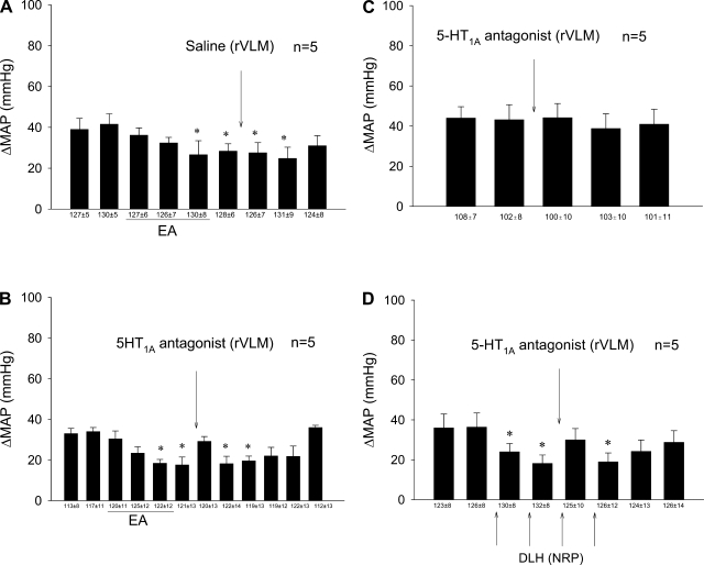
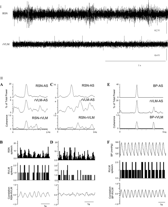
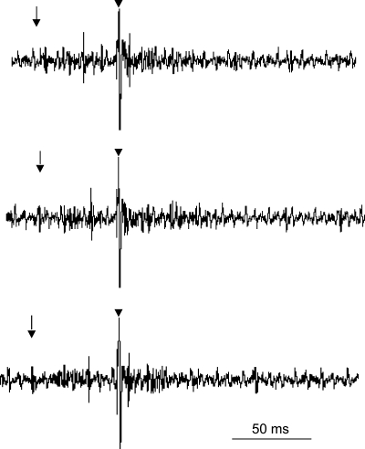
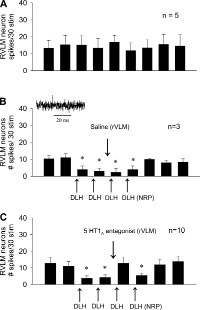
We have demonstrated that stimulation of somatic afferents during electroacupuncture (EA) inhibits sympathoexcitatory cardiovascular rostral ventrolateral medulla (rVLM) neurons and reflex responses. Furthermore, EA at P5-P6 acupoints over the median nerve on the forelimb activate serotonin (5-HT)-containing neurons in the nucleus raphe pallidus (NRP). The present study, therefore, examined the role of the NRP and its synaptic input to neurons in the rVLM during the modulatory influence of EA. Since serotonergic neurons in the NRP project to the rVLM, we hypothesized that the NRP facilitates EA inhibition of the cardiovascular sympathoexcitatory reflex response through activation of 5-HT1A receptors in the rVLM. Animals were anesthetized and ventilated, and heart rate and blood pressure were monitored. We then inserted microinjection and recording electrodes in the rVLM and NRP. Application of bradykinin (10 microg/ml) on the gallbladder every 10 min induced consistent excitatory cardiovascular reflex responses. Stimulation with EA at P5-P6 acupoints reduced the increase in blood pressure from 41+/-4 to 22+/-4 mmHg for more than 70 min. Inactivation of NRP with 50 nl of kainic acid (1 mM) reversed the EA-related inhibition of the cardiovascular reflex response. Similarly, blockade of 5-HT1A receptors with the antagonist WAY-100635 (1 mM, 75 nl) microinjected into the rVLM reversed the EA-evoked inhibition. In the absence of EA, NRP microinjection of dl-homocysteic acid (4 nM, 50 nl), to mimic EA, reduced the cardiovascular and rVLM neuronal excitatory reflex response during stimulation of the gallbladder and splanchnic nerve, respectively. Blockade of 5-HT1A receptors in the rVLM reversed the NRP dl-homocysteic acid inhibition of the cardiovascular and neuronal reflex responses. Thus activation of the NRP, through a mechanism involving serotonergic neurons and 5-HT1A receptors in the rVLM during somatic stimulation with EA, attenuates sympathoexcitatory cardiovascular reflexes.


Figures

Fig. 1.
Fig. 1.
Role of nucleus raphe pallidus (NRP) in electroacupuncture (EA) modulation of cardiovascular sympathoexcitatory reflex response. A and B: 30-min stimulation of P5-P6 overlying the median nerve caused prolonged attenuation of the increase in blood pressure (BP). B and C: depolarization blockade of the NRP with kainic acid (KA) reversed the inhibitory effects of EA. C: letters A–E above each BP tracing (top) represent bars (bottom). Bars represent increases (Δ) in mean arterial BP (MAP) induced by gallbladder simulation. BK, bradykinin. Means ± SE below histogram bars represent baseline MAP. *Significantly different compared with control MAP, P < 0.05.
Fig. 2.
Fig. 2.
Role of serotonin1A or 5-hydroxytryptamine (5-HT1A) receptors in rostral ventrolateral medulla (rVLM) during EA-cardiovascular modulation. A: 30-min stimulation of P5-P6 led to prolonged attenuation of the increase in BP in the presence of saline vehicle microinjection into the rVLM. B: the serotonin antagonist (WAY-100635), however, reversed the effects of EA. C: in contrast, the antagonist did not influence the cardiovascular responses in the absence of EA. D: microinjection of dl-homocysteic acid (DLH) into the NRP, like EA, caused sympathoinhibition, a response that was reversed by administration of the serotonin antagonist into the rVLM. Bars represent increases in MAP induced by gallbladder simulation. Means ± SE below histogram bars represent baseline MAP. *Significantly different compared with control MAP, P < 0.05.
Fig. 3.
Fig. 3.
Characterization of a cardiovascular sympathoexcitatory rVLM neuron that received convergent input from splanchnic nerve, median nerve, NRP, and baroreceptors. I (top): the activities of the renal sympathetic nerve (RSN) and the rVLM neuron are shown. II (bottom), A: the rVLM neuron displayed similar rhythmicity with renal sympathetic activity documented as a coherence of 0.6 at a frequency at 1.9 Hz and autospectra (AS). B: the neuron also demonstrated a close correlation in time, as measured with spike-triggered averaging. C and D: another rVLM cell (control neuron) and its correlation with renal sympathetic activity. Note lack of coherence and correlation. E: the same rVLM neuron discharged at an identical frequency of 1.9 Hz and demonstrated a coherence of 0.52 when its activity was gated with BP. F: the pulse-triggered averaging of BP and rVLM discharge frequency of this neuron. AS, ?.
Fig. 4.
Fig. 4.
Antidromic collision of a premotor rVLM neuron. The three panels display tracings that, from left to right, represent the splanchnic nerve-induced evoked spike, intermediolateral column (IML) stimulus, and antidromic-evoked action potential, respectively. The neuron had a conduction velocity of 11.5 m/s, a latency of 10 ms, and a refractory period of 7.6 ms. The middle tracing displays collision between the splanchnic nerve-induced orthodromic and the IML-evoked antidromic spikes. ↓, Time of stimulation of the splanchnic nerve; ↓, time of stimulation of the IML.
Fig. 5.
Fig. 5.
Role of 5-HT1A receptors in NRP-induced inhibition of rVLM neuronal activity. A: repeatable responses of premotor rVLM neurons following splanchnic nerve stimulation. B: DLH activation of the NRP neurons decreased discharge of sympathoexcitatory cardiovascular rVLM neurons. Inset: example of a rVLM spike. C: the serotonin antagonist (WAY-100635) reversed the effects of DLH-related inhibition of these rVLM neurons. *P < 0.05.
Fig. 6.
Fig. 6.
Composite map displaying sections of the medulla with sites of microinjections and recordings. Original pictures illustrate the sites (↓) of microinjection in the NRP (top) and recording in the rVLM (bottom). RFN, retrofacial nucleus; ION, inferior olivary nucleus; 5SP, alaminar spinal trigeminal nucleus.
Fig. 7.
Fig. 7.
Confocal microscopic images showing an NRP neuron double-labeled with c-Fos (A) and the retrograde microsphere tracer (B) injected into the rVLM of a rat (bregma −12.24 mm). C: merged image from A and B. Arrows in A, B, and C, respectively, indicate neurons containing c-Fos (green) and the retrograde tracer (red) and colocalization of c-Fos with the microsphere tracer. Scale bars in A–C represent 20 μm.

References

    1. Adams DB. The Behavioral and Brain Sciences. Brain Mechanisms for Offense, Defense and Submission. Cambridge, UK: Cambridge University Press, 1979, vol. 2
    1. Bago M, Dean C. Sympathoinhibition from ventrolateral periaqueductal gray mediated by 5-HT1A receptors in the RVLM. Am J Physiol Regul Integr Comp Physiol 280: R976–R984, 2001 - PubMed
    1. Bago M, Marson L, Dean C. Serotonergic projections to the rostroventrolateral medulla from midbrain and raphe nuclei. Brain Res 945: 249–258, 2002 - PubMed
    1. Barman SM, Gebber GL. Subgroups of rostral ventrolateral medullary and caudal medullary raphe neurons based on patterns of relationship to sympathetic nerve discharge and axonal projections. J Neurophysiol 77: 65–75, 1997 - PubMed
    1. Berman AL. The Brainstem of the Cat: A Cytoarchitectonic Atlas with Stereotaxic Coordinates. Madison, WI: University of Wisconsin Press, 1968

Publication types

MeSH terms