PPAR-gamma coactivator-1alpha regulates progesterone production in ovarian granulosa cells with SF-1 and LRH-1
- PMID: 20133449
- PMCID: PMC5419099
- DOI: 10.1210/me.2009-0352
PPAR-gamma coactivator-1alpha regulates progesterone production in ovarian granulosa cells with SF-1 and LRH-1
Abstract
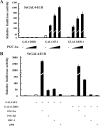
Previously, we demonstrated that bone marrow-derived mesenchymal stem cells (MSCs) differentiate into steroidogenic cells such as Leydig and adrenocortical cells by the introduction of steroidogenic factor-1 (SF-1) and treatment with cAMP. In this study, we employed the same approach to differentiate umbilical cord blood (UCB)-derived MSCs. Despite UCB-MSCs differentiating into steroidogenic cells, they exhibited characteristics of granulosa-luteal-like cells. We found that peroxisome proliferator-activated receptor-gamma coactivator-1alpha (PGC-1alpha) was expressed and further induced by cAMP stimulation in UCB-MSCs. Consistent with these results, tissue-specific expression of Pgc-1alpha was observed in rat ovarian granulosa cells. PGC-1alpha binds to the NR5A family [SF-1 and liver receptor homolog-1 (LRH-1)] of proteins and markedly enhances their transcriptional activities. Reporter assays revealed that PGC-1alpha activated the promoter activities of SF-1 and LRH-1 target genes. Infection of KGN cells (a human cell line derived from granulosa cells) with adenoviruses expressing PGC-1alpha resulted in the induction of steroidogenesis-related genes and stimulation of progesterone production. PGC-1alpha also induced SF-1 and LRH-1, with the latter induced to a greater extent. Knockdown of Pgc-1alpha in cultured rat granulosa cells resulted in attenuation of gene expression as well as progesterone production. Transactivation of the NR5A family by PGC-1alpha was repressed by Dax-1. PGC-1alpha binds to the activation function 2 domain of NR5A proteins via its consensus LXXLL motif. These results indicate that PGC-1alpha is involved in progesterone production in ovarian granulosa cells by potentiating transcriptional activities of the NR5A family proteins.
Figures


Similar articles
-
A novel isoform of liver receptor homolog-1 is regulated by steroidogenic factor-1 and the specificity protein family in ovarian granulosa cells.Endocrinology. 2013 Apr;154(4):1648-60. doi: 10.1210/en.2012-2008. Epub 2013 Mar 7. Endocrinology. 2013. PMID: 23471216
-
Nuclear receptor 5A (NR5A) family regulates 5-aminolevulinic acid synthase 1 (ALAS1) gene expression in steroidogenic cells.Endocrinology. 2012 Nov;153(11):5522-34. doi: 10.1210/en.2012-1334. Epub 2012 Sep 28. Endocrinology. 2012. PMID: 23024262
-
Liver receptor homolog-1 regulates the transcription of steroidogenic enzymes and induces the differentiation of mesenchymal stem cells into steroidogenic cells.Endocrinology. 2009 Aug;150(8):3885-93. doi: 10.1210/en.2008-1310. Epub 2009 Apr 9. Endocrinology. 2009. PMID: 19359379
-
The Orphan Nuclear Receptors Steroidogenic Factor-1 and Liver Receptor Homolog-1: Structure, Regulation, and Essential Roles in Mammalian Reproduction.Physiol Rev. 2019 Apr 1;99(2):1249-1279. doi: 10.1152/physrev.00019.2018. Physiol Rev. 2019. PMID: 30810078 Review.
-
Transcriptional regulation of genes related to progesterone production.Endocr J. 2015;62(9):757-63. doi: 10.1507/endocrj.EJ15-0260. Epub 2015 Jul 1. Endocr J. 2015. PMID: 26135521 Review.
Cited by
-
Differentiation of umbilical cord mesenchymal stem cells into steroidogenic cells in comparison to bone marrow mesenchymal stem cells.Cell Prolif. 2012 Apr;45(2):101-10. doi: 10.1111/j.1365-2184.2012.00809.x. Epub 2012 Feb 13. Cell Prolif. 2012. PMID: 22324479 Free PMC article.
-
The interaction of TRPV1 and lipids: Insights into lipid metabolism.Front Physiol. 2022 Dec 15;13:1066023. doi: 10.3389/fphys.2022.1066023. eCollection 2022. Front Physiol. 2022. PMID: 36589466 Free PMC article. Review.
-
Transcriptional Regulation of Ovarian Steroidogenic Genes: Recent Findings Obtained from Stem Cell-Derived Steroidogenic Cells.Biomed Res Int. 2019 Apr 1;2019:8973076. doi: 10.1155/2019/8973076. eCollection 2019. Biomed Res Int. 2019. PMID: 31058195 Free PMC article. Review.
-
Stem Leydig cells: from fetal to aged animals.Birth Defects Res C Embryo Today. 2010 Dec;90(4):272-83. doi: 10.1002/bdrc.20192. Birth Defects Res C Embryo Today. 2010. PMID: 21181888 Free PMC article. Review.
-
Diethylstilbestrol administration inhibits theca cell androgen and granulosa cell estrogen production in immature rat ovary.Sci Rep. 2017 Aug 21;7(1):8374. doi: 10.1038/s41598-017-08780-7. Sci Rep. 2017. PMID: 28827713 Free PMC article.
References
-
- Stocco C, Telleria C, Gibori G2007. The molecular control of corpus luteum formation, function, and regression. Endocr Rev 28:117–149 - PubMed
-
- Murphy BD2000. Models of luteinization. Biol Reprod 63:2–11 - PubMed
-
- Peng N, Kim JW, Rainey WE, Carr BR, Attia GR2003. The role of the orphan nuclear receptor, liver receptor homologue-1, in the regulation of human corpus luteum 3β-hydroxysteroid dehydrogenase type II. J Clin Endocrinol Metab 88:6020–6028 - PubMed
Publication types
MeSH terms
Substances
Grants and funding
LinkOut - more resources
Full Text Sources
Other Literature Sources
Molecular Biology Databases

