

Review
doi: 10.1126/science.1178331.
Development of monocytes, macrophages, and dendritic cells
Affiliations
- PMID: 20133564
- PMCID: PMC2887389
- DOI: 10.1126/science.1178331
Item in Clipboard
Review
Development of monocytes, macrophages, and dendritic cells
Science.
.
Erratum in
- Science. 2010 Dec 3;330(6009):1319
Abstract
Monocytes and macrophages are critical effectors and regulators of inflammation and the innate immune response, the immediate arm of the immune system. Dendritic cells initiate and regulate the highly pathogen-specific adaptive immune responses and are central to the development of immunologic memory and tolerance. Recent in vivo experimental approaches in the mouse have unveiled new aspects of the developmental and lineage relationships among these cell populations. Despite this, the origin and differentiation cues for many tissue macrophages, monocytes, and dendritic cell subsets in mice, and the corresponding cell populations in humans, remain to be elucidated.
Figures

(A). Still frames from time-lapse intravital confocal microscopy of a crawling monocytes (arrow) and perivascular macrophages in the dermis (courtesy of F. Geissmann, for details see (52)) (B). Confocal microscopy image of the spleen from mice grafted with MDPs. DCs derived from the MDP graft are labeled in green (CD45.2 staining) and host-derived marginal zone metallophilic macrophages are labelled in red with CD169/Sialoadhesin (courtesy of F. Geissmann, for details see (24)). (C). Dividing LCs in the epidermis (courtesy of I. Chorro & F. Geissmann, for details see (35)). (D). Confocal micrograph of aortic whole mount from Cx3cr1GFP/GFPApoe-/- mouse, viewed from endothelial side (courtesy of K. Ley). (E). Intra-vital two photon microscopic image of intestinal villi of CD11c+ cells-depleted mice reconstituted by grafts of bone marrow monocytes, yielding red and green fluorescent lamina propria cells, respectively (courtesy S. Jung and G. Shakhar, for details see (26)).
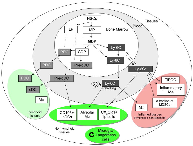
Differentiation of DCs and macrophages in mice. In the bone marrow, hematopoietic stem cells (HSC) produce myeloid (MP) and lymphoid (LP) committed precursors. MP give rise to monocyte/macrophages and DC precursors (MDP). MDP give rise to monocytes, and to common DC precursor (CDP). Two monocyte subsets, Ly-6C+ and Ly-6C- leave the bone marrow to enter the blood. CDP give rise to pre-classical dendritic cells (pre-cDC) and plasmacytoid dendritic cells (PDC). Pre-cDC circulate in blood and enter lymphoid tissue, where they give rise to CD8α+ and CD8α- cDCs, and non-lymphoid tissues, where they may give rise to CD103+ lamina propria DC (lpDC). Under homeostatic conditions, Ly-6C- monocytes may contribute to alveolar macrophages (MΦ) and Ly-6C+ monocytes can become CX3CR1+ lpDCs in non-lymphoid tissues. During inflammation, Ly-6C+ monocytes give rise to monocyte-derived DCs, e.g. TNF and iNOS-producing dendritic cells (TipDC), inflammatory macrophages, and may contribute to myeloid-derived suppressor cells (MDSC) associated with tumors. They are also suspected to contribute to microglia and Langerhans cells in selected experimental conditions. Microglia and Langerhans cells can renew independently from the bone marrow (curved arrow). HSC can also leave their bone marrow niche and enter peripheral tissues, where they differentiate to myeloid cells during inflammation. It is unclear at this time if LP contribute significantly to PDC and cDCs (dashed arrow).
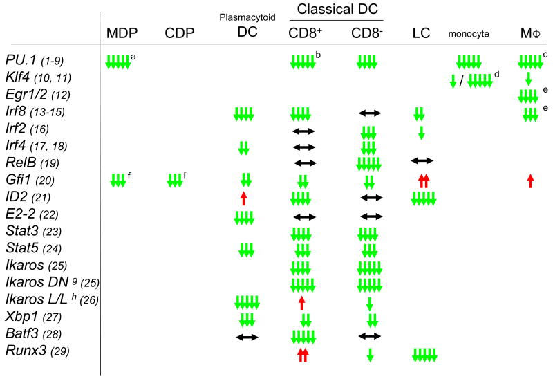
Phenotype of transcription factor knockout mice in different populations of the MPs. ↓ indicates a reduction in KO, ↑ an increase in KO and ↔ means unchanged numbers of the populations, suggesting a positive (green), negative (red) or no (black) effect on the development of the respective population. Number of arrows indicates the relative strength of the effect. Specific notes: (a) MDP have not been specifically analyzed but PU.1-deficient mice lack all myeloid progenitors of which MDP are a subpopulation, (b) in one study (SOM ref 7) CD8+ CD1 1c+ cells were detected in E16.5 embryos, (c) some macrophage subpopulations are present in the embryo, (d) LyC6-/LyC6+ respectively, (e) M-CSF dependent differentiation in culture, (f) MDP were not specifically studied, but the population of lin- ckit+ Flt3+ progenitors, which includes MDP was reduced; (g) dominant negative allele and (h) low level expression. References cited in this figure (1-29) are found in the SOM.
References
-
- Auffray C, Sieweke MH, Geissmann F. Annu Rev Immunol. 2009;27:669. - PubMed
Publication types
MeSH terms
Substances
Grants and funding
LinkOut - more resources
Full Text Sources
Other Literature Sources
