. 2010 Feb 5;327(5966):689-93.
doi: 10.1126/science.1181766.

Cryo-EM model of the bullet-shaped vesicular stomatitis virus

Affiliations

Cryo-EM model of the bullet-shaped vesicular stomatitis virus

Peng Ge et al. Science. .

Erratum in

  • Science. 2013 Feb 15;339(6121):757

Abstract

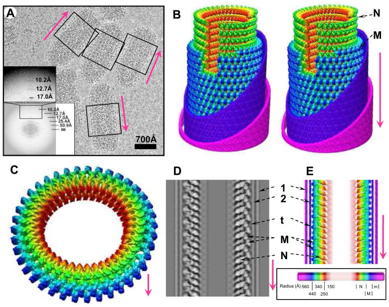
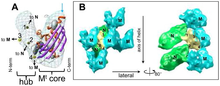
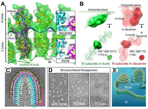
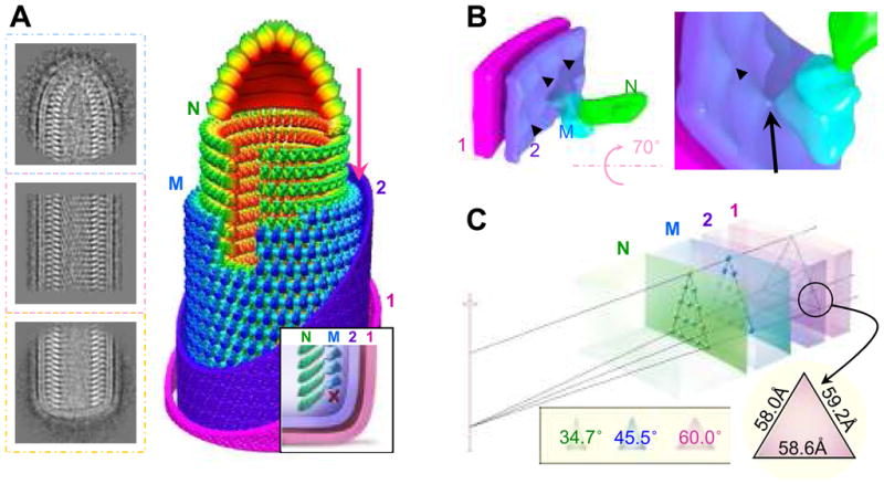
Vesicular stomatitis virus (VSV) is a bullet-shaped rhabdovirus and a model system of negative-strand RNA viruses. Through direct visualization by means of cryo-electron microscopy, we show that each virion contains two nested, left-handed helices: an outer helix of matrix protein M and an inner helix of nucleoprotein N and RNA. M has a hub domain with four contact sites that link to neighboring M and N subunits, providing rigidity by clamping adjacent turns of the nucleocapsid. Side-by-side interactions between neighboring N subunits are critical for the nucleocapsid to form a bullet shape, and structure-based mutagenesis results support this description. Together, our data suggest a mechanism of VSV assembly in which the nucleocapsid spirals from the tip to become the helical trunk, both subsequently framed and rigidified by the M layer.


Figures

Figure 1
Figure 1. CryoEM of VSV virion and 3D reconstruction of its helical trunk
(A) A typical cryoEM micrograph of VSV virions at 98,000x magnification. The trunk portion is marked by the boxes; inset, incoherent average of Fourier transforms of all raw images showing the layer lines. (B) Density map of the virion trunk. To enhance visual clarity and to show the interior, we computationally removed four turns of M, part of the membrane bilayer, incomplete subunits, and a 30° wedge. Nucleocapsid N and matrix M layers were displayed at a threshold of 1.15σ above the mean; envelope densities were displayed at a threshold of 0.1σ above the mean. (C) A complete repeat of the N and M helices, featuring 75 helical asymmetric units in two turns. (D) The central, vertical slice (17.3Å thick) in the density map and (E) a radially color-coded surface representation of a central slab (23Å thick). 1 and 2: outer and inner leaflets of the phospholipid bilayer envelope; t: putative cytoplasmic tail of G; M: matrix protein; N: nucleoprotein. Inset: Coloring scheme -- maps in all panels are colored according to radial distance as depicted in the scale bar. N: Nucleoprotein (red to green); M: Matrix protein (cyan to blue); e: envelope membrane (violet to pink). In this and the following figures, the arrow in every panel denotes the directionality (tip to trunk) of virions along the axis of the helix or of parts as they would be in the virion.
Figure 2
Figure 2. In situ structure of the full length M matrix protein
(A) Fit of the crystal structure (ribbon) of the C-terminal core domain MCTD (right part) of M into the corresponding density map (mesh, contoured at 1.15σ above the mean), taken from the cryoEM map. The α helices are shown in red and β sheets in purple. The numbered yellow spheres in the left part of the density map mark the positions of the four contact points on the “M-hub” domain. The highest density regions of the cryoEM density are shown as gray shaded surface by contouring at 3.0σ above the mean. (B) Left: four adjacent M (cyan) with two N (green) subunits in the neighborhood of one M with its M-hub in yellow. The contact points on the M-hub that mediate interactions with M and N are labeled 1–4. The volume is contoured at 1.0σ. Right: by turning the left panel 80° around the vertical axis and removing the frontal M subunit, the interaction between N and M is illustrated.
Figure 3
Figure 3. Formation of the “bullet” tip of VSV virion by the nucleocapsid (N) ribbon
(A) Fitting of the crystal structure (7) of nucleoprotein (N) (yellow ribbon) and RNA (blue ribbon) into the cryoEM density map (semi-transparent green, displayed at a threshold of 1.5σ above the mean) from the VSV virion trunk. The helical axis in this panel points toward the reader. The purple wire frames represents the highest density regions of the cryoEM structure (threshold of 3.5σ above the mean), which co-localize with α helices and the vRNA of the crystal structure. Insets: Along the upper part of the interface between adjacent C lobes in the decamer, there are six hydrogen bonds (including R309 to E419) and one (I237:Y324) hydrophobic interaction (top right inset). Following flexible docking of the atomic structure from the decamer into the cryoEM density map of the trunk, distances between amino acid partners in these seven sites increase by ~9Å, disrupting these interactions. (B) Comparison of the inclination of N subunits (green) in our cryoEM structure from the trunk of the virion (37.5 subunits/turn) with the inclination of the N subunits (red) in the crystal structure from the decamer ring (10 subunits/turn) (7). Top panels: Dashed lines through a side view of an N subunit from the trunk (left) and with an N subunit from the decamer (right) show the difference in tilt, the angle up from the horizontal plane. Bottom panels: Dashed lines through end-on views of N subunits show the difference in dihedral angle between adjacent N subunits in the trunk (green) and in the decamer ring (red). (C) A representative class-average of the virion tip from 75 individual images. Numbers inside the nucleocapsid designate the order of N subunits in the nucleocapsid ribbon, which may be traced by following the path 1 > 1′ > 2 > 2′, etc. (D) Negative stain electron microscopy images of the wild type decamer and two mutant rings confirm the importance of two of the interactions specified above. Both mutants produce rings larger than a decamer. (E) A cartoon illustration of a plausible process by which the nucleocapsid ribbon generates the virion head, starting with its bullet tip. The curling of the nucleocapsid ribbon generates a decamer-like turn at the beginning, similar to the crystal structure. When assembly nearly completes this turn, continuation of vRNA requires that the ribbon form a larger turn below it, similar to that in the mutants in (D). As the spiral enlarges and progresses to the helical trunk, the tilt of individual N subunits decreases. When it reaches the 7th turn, the nucleocapsid ribbon becomes helical (insets), where each new turn of the nucleocapsid fits naturally under the preceding turn (insets).
Figure 4
Figure 4. Architecture of the VSV virion and implication for pseudotyping
(A) Representative 2D averages of conical tip, trunk and base of VSV and a montage model of the tip and the cryoEM map of the trunk. N is green, M is blue, and the inner (“2”) and outer (“1”) leaflets of the membrane are violet and pink. Inset: illustration of the base region of the VSV virion. The “X” marks the absence of a turn of M helix below the lowest turn of the N helix. (B) CryoEM structure showing the putative cytoplasmic tail of G protein binding to an M subunit through a thin linker. The inner leaflet (“2”) of the membrane has unusual bumps (arrowhead) that meet M at the site of a thin linker density (arrow). (C) A wedge of the virion trunk, illustrating its geometric arrangement across the three layers. The N layer, the M layer, and the two membrane leaflets (“1” and “2”) are arranged in coaxial cylinders with their radii determined from our cryoEM structure. Due to the difference in radii, the helical lattice points on the three layers form different triangles. The smaller the radius, the narrower the apex angle (inset). At the outer surface of the membrane, the lattice points form an equilateral triangle.

Similar articles

Cited by

References

    1. Lichty BD, Power AT, Stojdl DF, Bell JC. Trends Mol Med. 2004 May;10:210. - PubMed
    1. Stojdl DF, et al. Cancer Cell. 2003 Oct;4:263. - PubMed
    1. Schnell MJ, Johnson JE, Buonocore L, Rose JK. Cell. 1997 Sep 5;90:849. - PubMed
    1. Mebatsion T, Finke S, Weiland F, Conzelmann KK. Cell. 1997 Sep 5;90:841. - PubMed
    1. Gaudier M, Gaudin Y, Knossow M. EMBO J. 2002 Jun 17;21:2886. - PMC - PubMed

Publication types

MeSH terms

Associated data