. 2010 Feb 23;107(8):3722-7.
doi: 10.1073/pnas.0915135107. Epub 2010 Feb 2.

Cancer stem cells from colorectal cancer-derived cell lines

Affiliations

Cancer stem cells from colorectal cancer-derived cell lines

Trevor M Yeung et al. Proc Natl Acad Sci U S A. .

Abstract

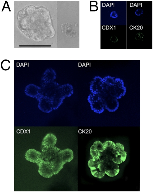
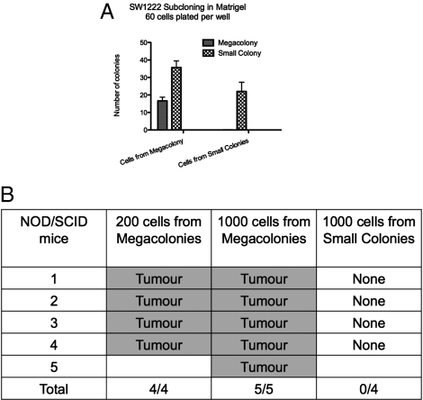
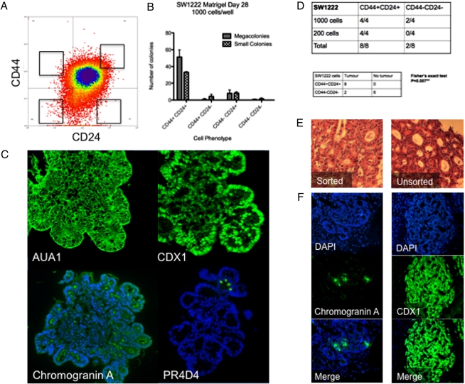
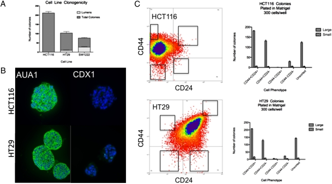
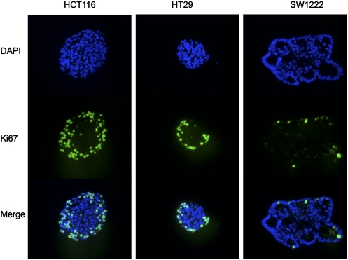
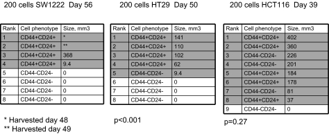
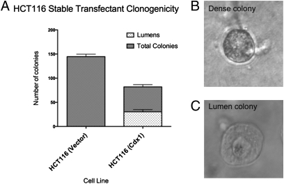
Cancer stem cells (CSCs) are the subpopulation of cells within a tumor that can self-renew, differentiate into multiple lineages, and drive tumor growth. Here we describe a two-pronged approach for the identification and characterization of CSCs from colorectal cancer cell lines, using a Matrigel-based differentiation assay, and cell surface markers CD44 and CD24. About 20 to 30% of cells from the SW1222 cell line form megacolonies in Matrigel that have complex 3D structures resembling colonic crypts. The megacolonies' capacity to self-renew in vitro is direct evidence that they contain the CSCs. Furthermore, just 200 cells from SW1222 megacolonies initiate tumors in NOD/SCID mice. We also showed that CD44(+)CD24(+) cells enriched for colorectal CSCs in the HT29 and SW1222 cell lines, which can self-renew and reform all four CD44/CD24 subpopulations, are the most clonogenic in vitro and can initiate tumors in vivo. A single SW1222 CD44(+)CD24(+) CSC, when grown in Matrigel, can form large megacolonies that differentiate into enterocyte, enteroendocrine, and goblet cell lineages. The HCT116 line does not differentiate or express CDX1, nor does it contain subpopulations of cells with greater tumor-forming capacity, suggesting that HCT116 contains mainly CSCs. However, forced expression of CDX1 in HCT116 leads to reduced clonogenicity and production of differentiating crypt-containing colonies, which can explain the selection for reduced CDX1 expression in many colorectal cancers. In summary, colorectal cancer cell lines contain subpopulations of CSCs, characterized by their cell surface markers and colony morphology, which can self-renew and differentiate into multiple lineages.


Conflict of interest statement

The authors declare no conflict of interest.

Figures

Fig. 1.
Fig. 1.
Immunofluorescence of SW1222 cell line colonies. (A) The SW1222 cell line can differentiate and form two distinct colony types: megacolonies with complex 3D lumen-like structures and small colonies. (Scale bar, 400 μm.) (B) Immunofluorescence of SW1222 megacolonies in Matrigel shows low levels of CDX1 and CK20 expression at day 7. (C) By day 14, megacolonies express much higher levels of CDX1 and CK20. Magnification: 20×.
Fig. 2.
Fig. 2.
SW1222 subcloning in Matrigel. (A) Megacolonies and small colonies growing in single wells in a 96-well plate were pooled separately, disaggregated into a single-cell suspension, and then replated in Matrigel in 96-well plates, at a concentration of 60 cells per well. Megacolonies are able to reform themselves, as well as small colonies, but small colonies can only reform themselves. (B) Cells from megacolonies are able to initiate tumors in NOD/SCID mice, but cells from small colonies are not tumorigenic.
Fig. 3.
Fig. 3.
Characterization of CD44/CD24 selected SW1222 cells. (A) The extreme 0.5 to 1% of each CD44/CD24 subgroup by FACS analysis was taken for further experiments. (B) CD44+CD24+ SW1222 cells are the most clonogenic and form the greatest numbers of megacolonies when grown in Matrigel, compared to other CD44/CD24 subgroups. (C) A single CD44+CD24+ cell can give rise to a megacolony forming multiple differentiated cell types: AUA-1 (anti-EpCam pan-epithelial marker), CDX1 (enterocyte), chromogranin A (enteroendocrine), and PR4D4 (mucin, see Fig. S6). Nuclei stained blue with DAPI. Megacolonies grown in Matrigel for 4 weeks. (D) CD44+CD24+ sorted cells are more tumorigenic in NOD/SCID mice: 200 and 1,000 cells of CD44+CD24+ and CD44CD24 cells were injected into NOD/SCID mice (Upper) and assessed for tumor forming capacity. The results of the 200 and 1,000 cell groups were combined together in a 2 × 2 contingency table (Lower), and analyzed statistically using Fisher’s exact test (P = 0.007). (E) Both CD44+CD24+ sorted and unsorted SW1222 cells are able to form well-differentiated adenocarcinomas that are histologically indistinguishable when injected into NOD/SCID mice (H&E stain, magnification 20×). (F) Xenografts derived from SW1222 CD44+CD24+ sorted cells express the differentiation markers chromogranin A (enteroendocrine) and CDX1 (enterocyte).
Fig. 4.
Fig. 4.
Cell line clonogenicity. (A) Clonogenicity of HCT116, HT29, and SW1222 in Matrigel. Numbers of lumen-forming and total colonies counted per well. One thousand cells plated in Matrigel per well in 96-well plates, grown for 2 weeks. (B) HCT116 and HT29 colonies grown in Matrigel for 2 weeks stain positive for AUA1. HCT116 colonies are CDX1-negative, and HT29 colonies weakly express CDX1. Nuclei stained blue with DAPI. Magnification, 20×. (C) The CD44+CD24+ and CD44+CD24 subpopulations are of comparable clonogenicity for HCT116 and HT29 cells grown in Matrigel. Three hundred cells seeded per well in 24-well plates.
Fig. 5.
Fig. 5.
Ki-67 staining for HCT116, HT29, and SW1222 colonies grown in Matrigel. Magnification, 20×.
Fig. 6.
Fig. 6.
Xenograft data ranked by size of tumor. SW1222 CD44+CD24+ cells are more tumourigenic than CD44CD24 cells (Fig. 3D). Tumors derived from HT29 CD44+CD24+ cells are significantly larger than those derived from CD44CD24 cells (unpaired t test, P < 0.001), but that is not the case for the HCT116 cell line (P = 0.27). Shaded rows highlight individual mice that developed tumors.
Fig. 7.
Fig. 7.
HCT116 stable-transfectant clonogenicity. (A) Forced CDX1 expression in HCT116 colonies reduces clonogenicity and results in the development of lumen-forming colonies. (B) HCT116 vector control cell line produces no lumen-forming colonies. (C) Forced CDX1 expression induces lumen formation within HCT116 colonies.

References

    1. Reya T, Morrison SJ, Clarke MF, Weissman IL. Stem cells, cancer, and cancer stem cells. Nature. 2001;414:105–111. - PubMed
    1. Bonnet D, Dick JE. Human acute myeloid leukemia is organized as a hierarchy that originates from a primitive hematopoietic cell. Nat Med. 1997;3:730–737. - PubMed
    1. O’Brien CA, Pollett A, Gallinger S, Dick JE. A human colon cancer cell capable of initiating tumour growth in immunodeficient mice. Nature. 2007;445:106–110. - PubMed
    1. Ricci-Vitiani L, et al. Identification and expansion of human colon-cancer-initiating cells. Nature. 2007;445:111–115. - PubMed
    1. Liu Y, Bodmer WF. Analysis of P53 mutations and their expression in 56 colorectal cancer cell lines. Proc Natl Acad Sci USA. 2006;103:976–981. - PMC - PubMed

Publication types