. 2010 Feb 23;107(8):3799-804.
doi: 10.1073/pnas.0914537107. Epub 2010 Feb 2.

Disruption of LGI1-linked synaptic complex causes abnormal synaptic transmission and epilepsy

Affiliations

Disruption of LGI1-linked synaptic complex causes abnormal synaptic transmission and epilepsy

Yuko Fukata et al. Proc Natl Acad Sci U S A. .

Abstract

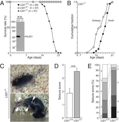
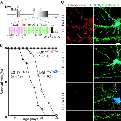
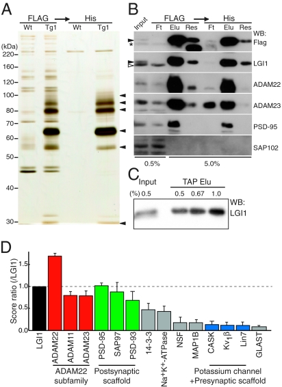
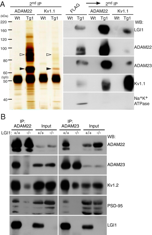
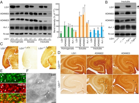
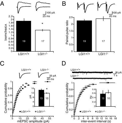
Epilepsy is a devastating and poorly understood disease. Mutations in a secreted neuronal protein, leucine-rich glioma inactivated 1 (LGI1), were reported in patients with an inherited form of human epilepsy, autosomal dominant partial epilepsy with auditory features (ADPEAF). Here, we report an essential role of LGI1 as an antiepileptogenic ligand. We find that loss of LGI1 in mice (LGI1(-/-)) causes lethal epilepsy, which is specifically rescued by the neuronal expression of LGI1 transgene, but not LGI3. Moreover, heterozygous mice for the LGI1 mutation (LGI1(+/-)) show lowered seizure thresholds. Extracellularly secreted LGI1 links two epilepsy-related receptors, ADAM22 and ADAM23, in the brain and organizes a transsynaptic protein complex that includes presynaptic potassium channels and postsynaptic AMPA receptor scaffolds. A lack of LGI1 disrupts this synaptic protein connection and selectively reduces AMPA receptor-mediated synaptic transmission in the hippocampus. Thus, LGI1 may serve as a major determinant of brain excitation, and the LGI1 gene-targeted mouse provides a good model for human epilepsy.


Conflict of interest statement

The authors declare no conflict of interest.

Figures

Fig. 1.
Fig. 1.
Loss of LGI1 causes lethal epilepsy in mice. (A) Survival plot of LGI1 mutant mice. The absence of LGI1 protein in the brain was confirmed with anti-LGI1 antibody (Inset). (B) Observed epileptic or lethal phenotype in LGI1−/− mice (n = 27) during the postnatal third week. (C) Epileptic behaviors of LGI1−/− mice at P17. The mutant animal had spontaneous generalized seizures (resulting in a blurred image; Upper), followed by full tonic extension (Lower; arrowheads indicate limb extension). (D) Response of 1-month-old wild-type (LGI1+/+) and LGI1+/− mice to PTZ (35 mg/kg). Data are mean ± SEM. Student’s t test: ***P < 0.001 (LGI1+/+, n = 12; LGI1+/−, n = 19). (E) Quantification of reaction to PTZ injection. Each criterion (0–5) is as follows. 0, no reaction; 1, twitching; 2, myoclonic body jerks; 3, clonic forelimb convulsions; 4, generalized clonic convulsions; 5, generalized tonic convulsions.
Fig. 2.
Fig. 2.
Neuronal expression of LGI1, but not LGI3, completely rescues the epileptic phenotype of LGI1−/− mice. (A) Thy1 promoter-driven LGI1 or LGI3 tagged with Flag and His × 6 (-FH) designed for generating transgenic mice. (B) Neuronal expression of LGI1-FH transgene (Tg1/+), but not LGI3 (Tg3/+), completely rescued the lethal epileptic phenotype of LGI1 knockout mice (LGI1−/−;+/+). (C) Thy1 promoter-driven LGI1-FH was functionally secreted and bound to surface-expressed ADAM22 (green) in cultured hippocampal neurons. LGI1 E383A-FH, an ADPEAF mutant, was not secreted and failed to bind to ADAM22. LGI3-FH did not show the binding to ADAM22-GFP–expressing neurons. Red, surface-bound LGIs-FH; blue, total expression of LGIs-FH (tLGI1). (Scale bars, 10 μm [Upper]; 5 μm [Lower; dendritic regions were magnified]).
Fig. 3.
Fig. 3.
Identification of global LGI1-containing protein complex in the brain. (A) In vivo LGI1-associated protein complex purified from the LGI1-FH–expressing mouse (Tg1), including p100, 90, 80, 65, 55, and 30 (arrowheads, from Top). (B) Major constituents of LGI1 complex contained ADAM23 as well as ADAM22 and PSD-95. Closed arrowheads, LGI1-FH; asterisk, nonspecific band; open arrowhead, endogenous LGI1; Ft, flow through; Elu, elution; Res, resin after elution. (C) More than 90% of expressed LGI1-FH were isolated by TAP. (D) LGI1-associated protein network analyzed by shotgun mass spectrometry. The identified specific proteins were lined up according to the SEQUEST score that is used for protein identification and semiquantification. Error bars, ±SD (n = 3).
Fig. 4.
Fig. 4.
LGI1 mediates the interaction between two receptors, ADAM22 and ADAM23. (A) LGI1, ADAM22, ADAM23, and Kv1 channel occur in a single protein complex. Purified LGI1 complex from the LGI1-FH expressing mouse was sequentially reprecipitated with antibodies to either ADAM22 or Kv1.1 (second IP). Open and closed arrowheads indicate ADAM22 and LGI1, respectively. (B) LGI1 connects between ADAM22 and ADAM23. ADAM22 (Left) or ADAM23 (Right) was immunoprecipitated from LGI1+/+ or LGI1−/− mouse brain extracts. Note that both immunoprecipitates includes postsynaptic PSD-95 and presynaptic Kv1.2.
Fig. 5.
Fig. 5.
Loss of LGI1/ADAM22/ADAM23 protein complex from the synapse causes epileptic phenotype. (A) Loss of LGI1 reduces ADAM22, ADAM23, and Kv1 channel in the synaptic fraction (insoluble). Crude synaptic membranes were fractionated by the Triton X-100 solubility, and probed with the indicated antibodies for quantitative Western blotting (Left). Relative band intensities compared with wild-type (+/+) samples are shown in graph. Deep color, LGI1+/−; light color, LGI1−/−. *P < 0.05; **P < 0.001. Error bars, ±SD (n = 3). (B) Reduction of synaptic ADAM22 and ADAM23 in LGI1−/− mouse was restored by the addition of an LGI1 transgene allele (LGI1−/−;Tg1/+) but not by an LGI3 allele (LGI1−/−;Tg3/+). Closed arrowheads, LGI1-FH; open arrowhead, endogenous LGI1. (C) LGI1 protein is highly enriched in the hippocampus and entorhinal cortex (transverse sections at P18). (Scale bar, 1 mm.) (D) Apparent reduction of ADAM22 and ADAM23 was observed in hippocampus of LGI1−/− mouse. (Scale bar, 0.2 mm.) (E) Immunofluorescence labeling of LGI1. LGI1 was often apposed to or colocalized with postsynaptic PSD-95 (arrowheads) at the molecular layer of the hippocampal regions. (Scale bar, 1 μm.) (F) Electronmicrography of hippocampal dentate gyrus shows that LGI1 localizes at the synaptic site (arrowheads). (Scale bar, 100 nm.)
Fig. 6.
Fig. 6.
LGI1 is essential for AMPAR-mediated synaptic currents. (A) Loss of LGI1 results in a significantly reduced (*P < 0.01) AMPA/NMDA ratio. (B) No difference in paired-pulse ratio is observed between wild-type (LGI1+/+) and LGI1−/− mice (P = 0.46). (C) Cumulative distribution plot and average amplitude, inset, showing a significant decrease (*P < 0.05) in mEPSC amplitude in LGI1−/− mice. (D) No change in frequency of mEPSCs was observed (P = 0.80).

Similar articles

Cited by

References

    1. Noebels JL. The biology of epilepsy genes. Annu Rev Neurosci. 2003;26:599–625. - PubMed
    1. Steinlein OK. Genetic mechanisms that underlie epilepsy. Nat Rev Neurosci. 2004;5:400–408. - PubMed
    1. Gu W, Brodtkorb E, Steinlein OK. LGI1 is mutated in familial temporal lobe epilepsy characterized by aphasic seizures. Ann Neurol. 2002;52:364–367. - PubMed
    1. Kalachikov S, et al. Mutations in LGI1 cause autosomal-dominant partial epilepsy with auditory features. Nat Genet. 2002;30:335–341. - PMC - PubMed
    1. Morante-Redolat JM, et al. Mutations in the LGI1/Epitempin gene on 10q24 cause autosomal dominant lateral temporal epilepsy. Hum Mol Genet. 2002;11:1119–1128. - PubMed

Publication types