. 2010 Feb 16;107(7):3157-62.
doi: 10.1073/pnas.0909587107. Epub 2010 Jan 28.

A broad-spectrum antiviral targeting entry of enveloped viruses

Affiliations

A broad-spectrum antiviral targeting entry of enveloped viruses

Mike C Wolf et al. Proc Natl Acad Sci U S A. .

Abstract

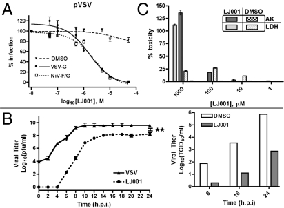
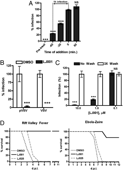
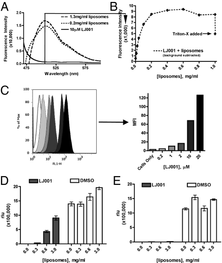
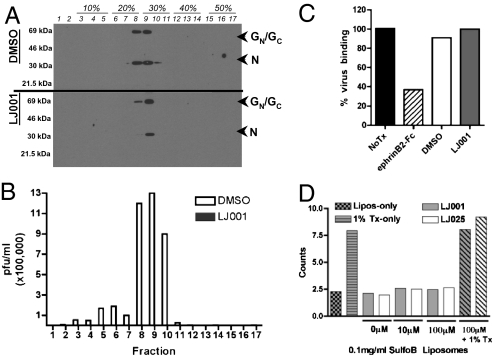
We describe an antiviral small molecule, LJ001, effective against numerous enveloped viruses including Influenza A, filoviruses, poxviruses, arenaviruses, bunyaviruses, paramyxoviruses, flaviviruses, and HIV-1. In sharp contrast, the compound had no effect on the infection of nonenveloped viruses. In vitro and in vivo assays showed no overt toxicity. LJ001 specifically intercalated into viral membranes, irreversibly inactivated virions while leaving functionally intact envelope proteins, and inhibited viral entry at a step after virus binding but before virus-cell fusion. LJ001 pretreatment also prevented virus-induced mortality from Ebola and Rift Valley fever viruses. Structure-activity relationship analyses of LJ001, a rhodanine derivative, implicated both the polar and nonpolar ends of LJ001 in its antiviral activity. LJ001 specifically inhibited virus-cell but not cell-cell fusion, and further studies with lipid biosynthesis inhibitors indicated that LJ001 exploits the therapeutic window that exists between static viral membranes and biogenic cellular membranes with reparative capacity. In sum, our data reveal a class of broad-spectrum antivirals effective against enveloped viruses that target the viral lipid membrane and compromises its ability to mediate virus-cell fusion.


Conflict of interest statement

The authors declare no conflict of interest.

Figures

Fig. 1.
Fig. 1.
Discovery of a broad-spectrum antiviral. (A) Pseudotyped VSV (pVSV) with the indicated envelope was pretreated with LJ001 or 0.1% DMSO (vehicle) for 10 min at 25 °C and then was used to infect Vero cells for 1 h at 37 °C (± SEM; normalized DMSO at 100%). (B) VSV-Indiana (Left) at an MOI of 3 was treated as in A, and infection was quantified by a standard plaque assay from supernatant samples (±SEM). **, P < 0.001 (>96% inhibition). NiV (Right) at an MOI of 3 was treated as in A with 10 μM LJ001, and measurements of the 50% tissue-culture infective dose were taken at the indicated time points. For both viruses in B, the infectious inoculum was replaced with growth media containing LJ001 at the indicated concentrations after the infection period indicated. (C) Vero cells were treated with varying concentrations of LJ001 for 1 h at 37 °C and assayed for lactate dehydrogenase (LDH) and adenylate kinase (AK) release (±SD). h.p.i., hours postinfection
Fig. 2.
Fig. 2.
LJ001 inactivates virions and prohibits viral entry. (A) pVSV was used to infect Vero cells as previously described. LJ001 (10 μM) was added at the indicated time relative to the infection end point (±SD). (B) Viruses were treated with 10 μM LJ001 for 10 min at 25 °C and then were washed with PBS, followed by repurification by ultracentrifugation through a sucrose cushion. Repurified viruses were used to infect cells as previously described (±SD). (C) Vero cells were treated with 1 μM or 10 μM compound for 10, 30, or 120 min at 37 °C in PBS (+10% FBS) and either left alone (No Wash) or washed three times (3 × Wash), followed by infection with pVSV (individual data sets normalized to corresponding vehicle control or negative compound, ±SD). (D) Equivalents of 100 × LD50 of RVFV-ZH501 or maZEBOV were treated ex vivo with 20 μM LJ001, 20 μM LJ025, or 2.5% DMSO for 20 min at 25 °C and then were used to infect mice (RVFV, n = 5; maZEBOV, n = 5) via i.p. injection. ***, P < 0.001; NS, not significant.
Fig. 3.
Fig. 3.
LJ001 binds, perturbs, and irreversibly targets the viral membrane. (A) Liposomes were titered into solution containing 10 μM LJ001 (excitation: 450 nm, emission: 510 nm), and fluorescence was monitored at the indicated wavelengths using a PTI QM4 fluorescence spectrophotometer (Perkin-Elmer). Representative raw data are shown. The solid line indicates no liposomes; dashed and dotted lines indicate increasing liposomal titrations. (B) A quantification of individual peaks at 510 nm as increasing concentrations of liposomes were titered into solution. Triton-X (0.1% final concentration) was added at the end of the assay to show that the increasing fluorescence depended on intact liposomes. These data were corrected for the scattering caused by the addition of liposomes by repeating the experiments in the absence of LJ001 and subtracting the liposome-induced scattering signal (±SEM). (C) (Left) Twenty-five thousand Vero cells were stained with increasing concentrations of LJ001 for 30 min at 37 °C in normal growth media and then were harvested by trypsinization and analyzed for mean fluorescence intensity (MFI) by flow cytometry. (Right) Bar graph shows MFI values. (D) Vero cells were infected with pVSV as previously described while simultaneously being subjected to 10 μM LJ001 and liposomes (±SD). (E) Vero cells were infected with pVSV as previously described. In this case, pVSV was treated with 10 μM LJ001 for 10 min at 25 °C and then subjected to varying concentrations of liposomes (±SD).
Fig. 4.
Fig. 4.
LJ001-treated virions remain grossly intact. (A) RVFV MP-12 was treated with 10 μM LJ001 or 2.5% DMSO for 20 min at 25 °C and banded across an iodixanol density gradient. One portion from each fraction was subjected to immunoblotting for the envelope (GC/GN) and nucleocapsid (N) proteins, and the other was used to conduct a plaque assay measuring infectivity. (B) Fractions from A were used to conduct a plaque assay measuring infectivity (white bars, DMSO; interleaved solid bars (not visible), LJ001). Note that the solid bars representing LJ001-treated viruses cannot be seen in the figure and represent at least a 5-log reduction in infectivity. Similar results were obtained upon repetition with RVFV as well as pVSV. (C) CHO cells stably expressing ephrinB2 were incubated with NiV-pVSV in the presence of 0.1% DMSO, 10 μM LJ001, or 40 nM soluble ephrin B2-Fc (EFN-B2) at 4 °C for 2 h. Cells were washed and fixed in 0.5% PFA; then bound viruses were detected with anti-NiV-F and quantified by flow cytometry. This panel is a graphical representation of raw histogram data from a representative experiment. (D) Sulforhodamine B-loaded liposomes (200 nm) were incubated with the indicated concentration of compound and assayed for fluorescent signal. Data were collected using a PTI QM4 fluorescence spectrophotometer at 25 °C (with constant stirring) with 4-nm excitation/emission bandpass at 560-nm excitation and 582-nm emission (counts ×100,000).
Fig. 5.
Fig. 5.
LJ001 affects viral–cell fusion but not cell–cell fusion. (A) NiV virus-like particles, pretreated with 10 μM LJ001 or DMSO, were used to infect Vero cells preloaded with CCF2-AM substrate and assayed for infection via flow cytometry (32). Data are shown as normalized ratios of blue:green cells (±SEM). (B) Vero cells were transfected with NiV-F and -G expression vectors, incubated overnight in media with 10 μM LJ001 or 0.1% DMSO, DAPI stained, and assayed visually for nuclei in syncytia by counting and averaging five 10× fields (±SD). (C) The treatment of cells with the fatty acid synthesis inhibitor TOFA increases relative toxicity of LJ001-treated cells. Vero cells were incubated with media containing the indicated concentration of TOFA in the presence of the indicated concentration of LJ compound for 24 h and then were measured for cellular toxicity using the Toxilight assay (Cambrex). Data represent n = four per goup in triplicate experiments. Data are normalized to 100% toxicity (indicated by 100% cell lysis) with toxicity of the indicated TOFA concentration subtracted as background. *, P < 0.05; **, P < 0.01; ***, P < 0.001; NS, not significant.

Comment in

Similar articles

Cited by

References

    1. Tam RC, Lau JY, Hong Z. Mechanisms of action of ribavirin in antiviral therapies. Antivir Chem Chemother. 2001;12:261–272. - PubMed
    1. Bekisz J, Schmeisser H, Hernandez J, Goldman ND, Zoon KC. Human interferons alpha, beta and omega. Growth Factors. 2004;22:243–251. - PubMed
    1. de Veer MJ, et al. Functional classification of interferon-stimulated genes identified using microarrays. J Leukoc Biol. 2001;69:912–920. - PubMed
    1. Sen GC. Viruses and interferons. Annu Rev Microbiol. 2001;55:255–281. - PubMed
    1. Hong Z, Cameron CE. Pleiotropic mechanisms of ribavirin antiviral activities. Prog Drug Res. 2002;59:41–69. - PubMed

Publication types