Comparative Study
. 2010 Feb 16;107(7):3006-11.
doi: 10.1073/pnas.0914839107. Epub 2010 Jan 28.

Analysis of CD161 expression on human CD8+ T cells defines a distinct functional subset with tissue-homing properties

Affiliations
Comparative Study

Analysis of CD161 expression on human CD8+ T cells defines a distinct functional subset with tissue-homing properties

Eva Billerbeck et al. Proc Natl Acad Sci U S A. .

Abstract

CD8(+) T lymphocytes play a key role in host defense, in particular against important persistent viruses, although the critical functional properties of such cells in tissue are not fully defined. We have previously observed that CD8(+) T cells specific for tissue-localized viruses such as hepatitis C virus express high levels of the C-type lectin CD161. To explore the significance of this, we examined CD8(+)CD161(+) T cells in healthy donors and those with hepatitis C virus and defined a population of CD8(+) T cells with distinct homing and functional properties. These cells express high levels of CD161 and a pattern of molecules consistent with type 17 differentiation, including cytokines (e.g., IL-17, IL-22), transcription factors (e.g., retinoic acid-related orphan receptor gamma-t, P = 6 x 10(-9); RUNX2, P = 0.004), cytokine receptors (e.g., IL-23R, P = 2 x 10(-7); IL-18 receptor, P = 4 x 10(-6)), and chemokine receptors (e.g., CCR6, P = 3 x 10(-8); CXCR6, P = 3 x 10(-7); CCR2, P = 4 x 10(-7)). CD161(+)CD8(+) T cells were markedly enriched in tissue samples and coexpressed IL-17 with high levels of IFN-gamma and/or IL-22. The levels of polyfunctional cells in tissue was most marked in those with mild disease (P = 0.0006). These data define a T cell lineage that is present already in cord blood and represents as many as one in six circulating CD8(+) T cells in normal humans and a substantial fraction of tissue-infiltrating CD8(+) T cells in chronic inflammation. Such cells play a role in the pathogenesis of chronic hepatitis and arthritis and potentially in other infectious and inflammatory diseases of man.


Conflict of interest statement

The authors declare no conflict of interest.

Figures
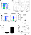
Fig. 1.
Fig. 1.
Function of CD161-expressing CD8+ T cells. (A) FACS profile of CD161 expression on CD3+CD8+ T cells in healthy donors. Cells within the live lymphocyte CD3+ gate are shown. The gates shown here were also the gates used for sorting (B). Panels show PMA/ionomycin–stimulated expression of IFN-γ (Upper) and TNF-α (Lower) according to CD161 expression, using gates as indicated in Fig 1A. Unstimulated controls are shown in Fig. S1D. (C) IL-17 production in PMA/ionomycin–stimulated cells (gated as in Fig 1A). (D) Group data from 10 healthy control subjects showing percentage of IL-17 production in CD161++, CD161+, and CD161 CD8+ T cells. (E). Secretion of IL-17 as measured by ELISA after stimulation of sorted cells. ICS is shown below for comparison. One representative experiment of two is shown. (F) Expression of IL-22 in healthy donors. Healthy donor PBMCs were stimulated as in Fig 1B and IL-22 secretion determined in CD161++ cells. The percentage of cells staining positive is indicated.
Fig. 2.
Fig. 2.
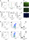
Phenotype of CD161++ cells (A) Relative expression of levels of RORC (RORγt) in CD161, CD161+, and CD161++ sorted CD8+ T cells (shown as , +, and ++). Data are derived from the RNA expression analyses after normalization among the 12 samples (n = 4 per group). (B) Expression of RORγT by flow cytometry in CD161++, CD161+, and CD161 CD3+CD8+ T cells. Representative histogram plot (Left) and combined data (Right); *P = 0.002. (C) Expression of RORγT by immunofluorescence of sorted CD161++ cells (Upper) and CD161 cells (Lower); DAPI staining is shown for CD161 cells. Magnification: ×200. (D) Expression of IL23R by gene array (Left), qRT-PCR (Center), and flow cytometry (Right). Combined staining from six healthy donors is shown. (E) IL-18 receptor (IL18R) expression on CD161++ cells: gene expression (Left), example flow cytometry (Center), and group flow cytometry data (Right). *P < 0.0001. (F) CCR2 expression on CD161++ cells displayed as in Fig 1E.
Fig. 3.
Fig. 3.
Peripheral and intrahepatic distribution of IL-17–secreting CD8+ T cells during chronic HCV infection. (A) Analysis of intracellular production of IL-17 and IFN-γ after stimulation with PMA/ionomycin in CD8+ T cells expanded from PBMCs and livers of HCV-infected patients. (B) FACS plot from one representative subject showing the distribution of IL-17– and IFN-γ–secreting CD8+ T cells in blood and liver. (C) Representative FACS plot showing CD161 expression on peripheral and intrahepatic IL-17–secreting CD8+ T cells. (D) Polyfunctionality of IL17+ cells: Percentages of IL-17–secreting, IL-17/IFN-γ–secreting, IL-17/IL-22–secreting, and IL-17/IL-22/IFN-γ–secreting cells among the liver and blood-derived IL-17–producing CD8+ T cell populations.
Fig. 4.
Fig. 4.
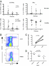
Frequencies of tissue-homing TC17 cells. (A) Correlations between the frequency of intrahepatic IFN-γ– or IL-17/IFN-γ–producing CD8+ T cells and clinicopathologic disease score (Metavir) in HCV patients. Increasing Metavir score relates to increasing levels of intrahepatic fibrosis, with 4 being the most severe (i.e., cirrhosis). Each dot represents one patient. (Upper) Cytokines are analyzed on ex vivo–derived cells (using PMA/ionomycin stimulation); (Lower) Cells are analyzed in a separate set of patients after nonspecific in vitro expansion followed by PMA/ionomycin stimulation as in Fig 3 (Spearman correlation). (B) CD161-expressing CD8+ T cells from an inflamed joint secreting IL-17 after PMA/ionomycin stimulation. One representative example (psoriatic arthritis) is shown (PBMCs below). (C) Group data from four donors (n = 2 psoriatic, n = 1 rheumatoid, n = 1 reactive arthritis; paired t test). (D) Relative secretion of IL-17 in CD161++ versus CD161+/− cells in joints (Mann-Whitney test for ++ vs. +/).

Similar articles

Cited by

References

    1. Klenerman P, Hill A. T cells and viral persistence: lessons from diverse infections. Nat Immunol. 2005;6:873–879. - PubMed
    1. Appay V, et al. Memory CD8+ T cells vary in differentiation phenotype in different persistent virus infections. Nat Med. 2002;8:379–385. - PubMed
    1. Lauer GM, Walker BD. Hepatitis C virus infection. N Engl J Med. 2001;345:41–52. - PubMed
    1. Bowen DG, Walker CM. Adaptive immune responses in acute and chronic hepatitis C virus infection. Nature. 2005;436:946–952. - PubMed
    1. He XS, et al. Quantitative analysis of hepatitis C virus-specific CD8(+) T cells in peripheral blood and liver using peptide-MHC tetramers. Proc Natl Acad Sci USA. 1999;96:5692–5697. - PMC - PubMed

Publication types

MeSH terms