. 2010 Feb 16;107(7):3163-8.
doi: 10.1073/pnas.0911979107. Epub 2010 Jan 28.

Bacterial translation elongation factor EF-Tu interacts and colocalizes with actin-like MreB protein

Affiliations

Bacterial translation elongation factor EF-Tu interacts and colocalizes with actin-like MreB protein

Hervé Joël Defeu Soufo et al. Proc Natl Acad Sci U S A. .

Abstract

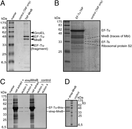
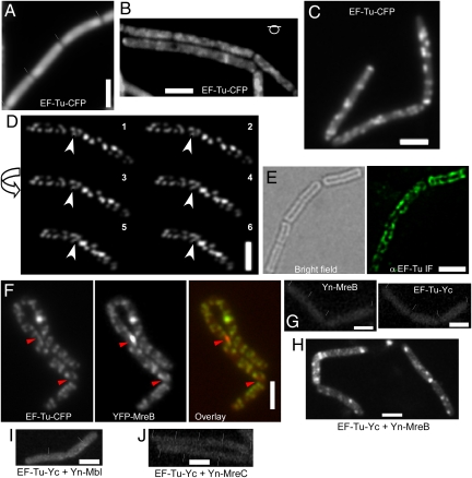
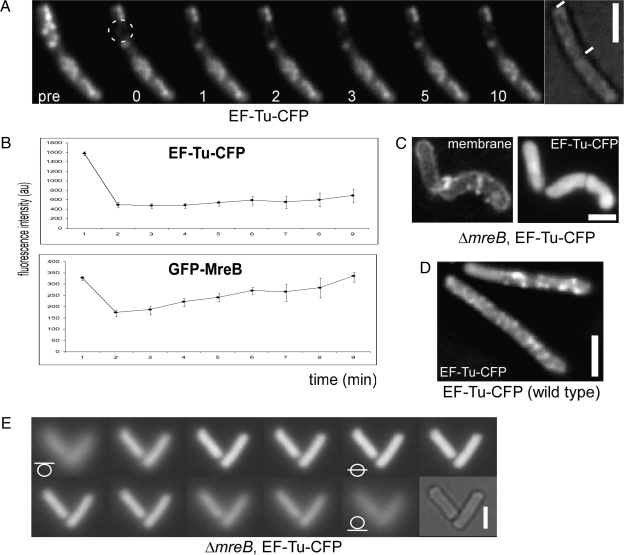
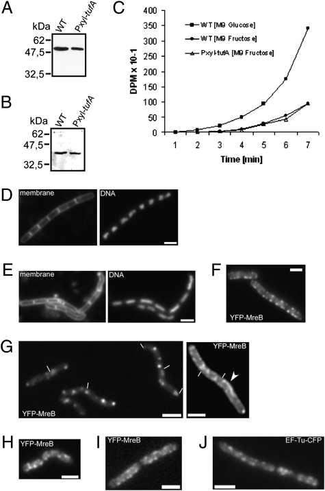
We show that translation initiation factor EF-Tu plays a second important role in cell shape maintenance in the bacterium Bacillus subtilis. EF-Tu localizes in a helical pattern underneath the cell membrane and colocalizes with MreB, an actin-like cytoskeletal element setting up rod cell shape. The localization of MreB and of EF-Tu is interdependent, but in contrast to the dynamic MreB filaments, EF-Tu structures are more static and may serve as tracks for MreB filaments. In agreement with this idea, EF-Tu and MreB interact in vivo and in vitro. Lowering of the EF-Tu levels had a minor effect on translation but a strong effect on cell shape and on the localization of MreB, and blocking of the function of EF-Tu in translation did not interfere with the localization of MreB, showing that, directly or indirectly, EF-Tu affects the cytoskeletal MreB structure and thus serves two important functions in a bacterium.


Conflict of interest statement

The authors declare no conflict of interest.

Figures

Fig. 1.
Fig. 1.
Coomassie-stained SDS/PAGE experiments showing the interaction of MreB and EF-Tu. The identities of coeluting bands are indicated by triangles or lines. (A) TAP-tag experiment of cells expressing TAP-MreB (JS82); shown are the elution fractions of the second affinity column. Control, extracts from cells expressing TAP-tag only; TAP-MreB, extracts from cells expressing TAP-MreB. (B) TAP-tag experiments of cells expressing EF-Tu-TAP or TAP-tag only (“control”); shown are second elution fractions. (C) Strep-MreB and EF-Tu-6His coelution from streptavidin columns: overexpression of both proteins (“strep-MreB”) and or just of EF-Tu-6His (“control”) in E. coli cells. Shown are main MreB elution fractions. (D) Coelution of purified EF-Tu-6His from Streptavidin columns preloaded with purified strep-MreB (“strep-MreB”) or with elution fractions from a Streptavidin column from E. coli cell extract lacking any overexpressed protein (“control”).
Fig. 2.
Fig. 2.
Localization of EF-Tu in B. subtilis cells. (AC) Cells expressing EF-Tu-CFP in B. Subtilis (JS88) at the original locus early exponential phase (A) (septa between cells are indicated by white lines and can usually be seen due to a lack of fluorescence between the cells), mid exponential phase (B), late exponential phase (C). (D) Three-dimensional deconvolution images. Arrow, direction of the turning angle of the cell; white arrowhead, an apparent helix. (E) Immunofluorescence on B. subtilis wild-type cells (late exponential growth) using anti-EF-Tu antiserum. (F) Two-dimensional deconvolution images showing colocalization of YFP-MreB and EF-Tu-CFP (strain JS89, late exponential growth). Red arrowheads, YFP-MreB foci (red in the overlay) that do not colocalize with EF-Tu-CFP foci (green in the overlay image). (GJ) BiFC interaction studies. Yn, N-terminal part of the split YFP; Yc, C-terminal part [note that coexpression of EF-Tu-Yc and just the Yn fragment (JS92) or of Yn-MreB and just the Yc fragment (JS93) did not show any defined signal]. (G) Expression of Yn-MreB (JS71; Left) or of EF-Tu-Yc (JS90; Right). (H) Cells expressing both EF-Tu-Yc and Yn-MreB (JS94). (I) Cells expressing of both EF-Tu-Yc and Yn-Mbl (JS95). (J) Cells expressing both EF-Tu-Yc and Yn-MreC (JS96). Cells were grown in S750 minimal media at room temperature supplemented with necessary antibiotics or inducers (0.5% xylose for YFP-MreB or 1 mM IPTG for Yn-fusions). (Scale bars: 2 μm.)
Fig. 3.
Fig. 3.
Dynamics of EF-Tu-CFP filaments and effect of the loss of MreB. (A) FRAP experiment on EF-Tu-CFP-expressing cells (JS88). pre, before bleaching; dotted circle, area of bleaching; numbers, minutes after bleaching. (B) FRAP profiles of EF-Tu-CFP (JS88) and of GFP-MreB (JS12). (C) Two-dimensional deconvoluted images of the localization of EF-Tu-CFP in mreB null cells (JS97). (D) Two-dimensional deconvoluted image of EF-Tu-CFP in wild-type cells (JS88). (E) Z-stack through mreB mutant cells expressing EF-Tu-CFP (JS97). White lines through circles, position of the focal plane. For FRAP experiments, strains JS12 and JS88 were grown in S750 minimal media at room temperature supplemented with necessary antibiotics or inducers (0.5% xylose for GFP-MreB). Strains JS88 and JS97 were grown in PAB/SMM medium (high magnesium and sucrose concentration). Late exponentially growing cells (OD 1.5–3) were used for the microscopy. (Scale bars, 2 μm.)
Fig. 4.
Fig. 4.
Effect of the depletion of EF-Tu in B. subtilis cells. (A) Western blot using EF-Tu antiserum showing levels of EF-Tu in wild-type cells (WT) and in the pxyl-tufA strain (JS91, OD = 0.5, M9 minimal medium plus fructose and xylose). (B) Western blot using MreB antiserum showing levels of MreB in wild-type (WT) cells and in the pxyl-tufA strain (JS91, OD = 0.5). (C) Translation efficiency (incorporation of radioactive methionine into cellular proteins) in wild-type and pxyl-tufA (JS91) cells. Cells were grown in M9 medium with fructose and xylose until early exponential phase (OD600 = 0.5), where wild-type and JS91 cells showed the same doubling time (Fig. S3). See SI Materials and Methods for details. (DG) Fluorescence microscopy of early exponential phase (OD600 = 0.5) growing B. subtilis cells in M9 medium with fructose and xylose. (D) Wild-type cells. (E) pxyl-tufA cells (JS91, EF-Tu depletion). (F) Localization of YFP-MreB in wild-type cells (JS36). (G) pxyl-tufA cells (JS98). Arrowhead, irregular nonhelical filament; white lines, ends of cells. (H) Cells of strain JS98 (YFP-MreB, pxyl-tufA) growing in SMM/PAB medium (high magnesium and sucrose). (IJ) Cells 1 h after addition of kirromycin, where growth had ceased, after growth in S750 minimal medium to late exponential phase. (I) Cells expressing YFP-MreB (JS36). (J) Cells expressing EF-Tu-CFP (JS88). (Scale bars, 2 μm.)

Similar articles

Cited by

References

    1. Matsuzawa H, Hayakawa K, Sato T, Imahori K. Characterization and genetic analysis of a mutant of Escherichia coli K-12 with rounded morphology. J Bacteriol. 1973;115:436–442. - PMC - PubMed
    1. Wachi M, Doi M, Okada Y, Matsuhashi M. New mre genes mreC and mreD, responsible for formation of the rod shape of Escherichia coli cells. J Bacteriol. 1989;171:6511–6516. - PMC - PubMed
    1. Bendezú FO, Hale CA, Bernhardt TG, de Boer PA. RodZ (YfgA) is required for proper assembly of the MreB actin cytoskeleton and cell shape in E. coli. EMBO J. 2009;28:193–204. - PMC - PubMed
    1. Alyahya SA, et al. RodZ, a component of the bacterial core morphogenic apparatus. Proc Natl Acad Sci USA. 2009;106:1239–1244. - PMC - PubMed
    1. Shiomi D, Sakai M, Niki H. Determination of bacterial rod shape by a novel cytoskeletal membrane protein. EMBO J. 2008;27:3081–3091. - PMC - PubMed

Publication types

MeSH terms

LinkOut - more resources