. 2010 Feb 16;107(7):3064-9.
doi: 10.1073/pnas.0912393107. Epub 2010 Jan 28.

Premature replacement of mu with alpha immunoglobulin chains impairs lymphopoiesis and mucosal homing but promotes plasma cell maturation

Affiliations

Premature replacement of mu with alpha immunoglobulin chains impairs lymphopoiesis and mucosal homing but promotes plasma cell maturation

Sophie Duchez et al. Proc Natl Acad Sci U S A. .

Abstract

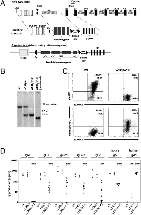
Sequentially along B cell differentiation, the different classes of membrane Ig heavy chains associate with the Ig alpha/Ig beta heterodimer within the B cell receptor (BCR). Whether each Ig class conveys specific signals adapted to the corresponding differentiation stage remains debated. We investigated the impact of the forced expression of an IgA-class receptor throughout murine B cell differentiation by knocking in the human C alpha Ig gene in place of the S mu region. Despite expression of a functional BCR, homozygous mutant mice showed a partial developmental blockade at the pro-B/pre-BI and large pre-BII cell stages, with decreased numbers of small pre-BII cells. Beyond this stage, peripheral B cell compartments of reduced size developed and allowed specific antibody responses, whereas mature cells showed constitutive activation and a strong commitment to plasma cell differentiation. Secreted IgA correctly assembled into polymers, associated with the murine J chain, and was transported into secretions. In heterozygous mutants, cells expressing the IgA allele competed poorly with those expressing IgM from the wild-type allele and were almost undetectable among peripheral B lymphocytes, notably in gut-associated lymphoid tissues. Our data indicate that the IgM BCR is more efficient in driving early B cell education and in mucosal site targeting, whereas the IgA BCR appears particularly suited to promoting activation and differentiation of effector plasma cells.


Conflict of interest statement

The authors declare no conflict of interest.

Figures

Fig. 1.
Fig. 1.
IgA1 expression instead of IgM in α1KI mice. (A) (Top) Structure of the targeted locus (not to scale) showing an unrearranged IgH locus and the extent of the deletion within the Iμ-Cμ intron. (Middle) Structure of the targeting vector in which Cα1 and a neor cassette flanked by loxP sites were inserted in place of Sμ. (Bottom) The α1KI locus is able to undergo V(D)J rearrangement after insertion of Cα1, whereas the neo gene can be deleted by cre-mediated recombination. (B) Southern blot analysis of tail DNA from representative WT, heterozygous, homozygous, and neo-deleted mutant mice. (C) Splenocytes from WT (Left) or α1KI/α1KI mice (Right) were labeled with PC5-conjugated anti-B220 and with either FITC-conjugated anti-IgM Abs showing the blockade of the IgM BCR expression in mutant animals (Top) or FITC-conjugated anti-human IgA1 Abs (Bottom). (D) Total endogenous Ig production was estimated by ELISA in sera from 6-week-old WT, α1KI/+, or α1KI/α1KI mice. Asterisks mark statistically significant differences with controls (Student’s t test, **P < 0.01; ***P < 0.001). The vertical axis is logarithmic; values are indicated as μg/mL.
Fig. 2.
Fig. 2.
Global in vivo increase of the plasma cell compartment in mutant mice. (A and B) Staining of tissues from WT (Left) and α1KI/α1KI mice (Right) with hemalun and revelation of human IgA1 with HRP-conjugated Ig show the presence of plasma cells (arrows) producing IgA1 and accumulating in (A) the splenic marginal zone and red pulp or (B) around Peyer's patches and in the lamina propria along intestinal crypts of the jejunum. (Original magnification, 100×.) (C) Staining of all plasma cells with anti-mouse Ig (HC + LC) shows accumulation of plasma cells in the spleen of a mutant mouse by comparison with WT. (Original magnification, 200×.) (D) Staining for CD138 and intracellular κ LC shows ASCs as CD138+ cells brightly staining for κ. (E) Staining for CD138+ and intracellular κ LC+ ASCs in splenocytes from SLP65-deficient mice. (F) Absence of significant difference by cell flow cytometry between proportions of recently differentiated (CD138+BrdU+) versus long-lived plasma cells (CD138+BrdU−) from α1KI/α1KI and WT spleens (means from three and four animals fed with BrdU, respectively). (G) Expression of Blimp-1 transcript in lymphoid tissues by qPCR evaluates the amount of cells engaged in plasma cell differentiation among all of the cells present in tissue samples (reference to 18S RNA) or by comparison with the pool of B lymphocytes (CD79a as a reference transcript). Total RNA was prepared without prior immunization of (▲) WT and (•) α1KI/α1KI mice or 3 days after intraperitoneal immunization of (∆) WT and (○) α1KI/α1KI mice with BSA.
Fig. 3.
Fig. 3.
Mutant B cells are precommitted to plasma cell differentiation. (A) Proliferation of sorted CD43− splenocytes (standardized on the numbers of CD19+ B cells) from α1KI/α1KI mice compared to WT (gray) after 4 days in vitro stimulation by LPS (Top), anti-CD40 plus IL-4 (Middle), or anti-κ Abs plus IL-4 (Bottom). (B) Ratios of CD138- versus CD19-expressing cells compared between WT (gray) and α1KI/α1KI mice within splenocytes stimulated for 2 days by LPS, anti-CD40 plus IL-4, or anti-κ LC plus IL-4 (*P < 0.05; **P < 0.01; ***P < 0.001 significant difference).
Fig. 4.
Fig. 4.
Confocal imaging of antibody-secreting cells in WT and mutant tissues. Evaluation of the number of ASCs secreting human IgA1 (green) versus IgM (red) by confocal microscopy in WT (Left), α1KI/α1KI (Center), or α1KI/+ heterozygous mice bred in an AID−/− background (Right). In the latter animals, counting of plasma cells in several microscopic fields showed that 24 out of 350 plasma cells (i.e., 7.4%) in the spleen were producing IgA1, whereas there was no IgA1-positive cell detectable in gut-associated lymphoid tissues. (Original magnification, 630×.)

References

    1. Brandtzaeg P. Induction of secretory immunity and memory at mucosal surfaces. Vaccine. 2007;25:5467–5484. - PubMed
    1. Kerr MA. The structure and function of human IgA. Biochem J. 1990;271:285–296. - PMC - PubMed
    1. Flanagan JG, Lefranc MP, Rabbitts TH. Mechanisms of divergence and convergence of the human immunoglobulin α 1 and α 2 constant region gene sequences. Cell. 1984;36:681–688. - PubMed
    1. Word CJ, Mushinski JF, Tucker PW. The murine immunoglobulin α gene expresses multiple transcripts from a unique membrane exon. EMBO J. 1983;2:887–898. - PMC - PubMed
    1. Cogné M, Preud’homme JL. Gene deletions force nonsecretory α-chain disease plasma cells to produce membrane-form α-chain only. J Immunol. 1990;145:2455–2458. - PubMed

Publication types

MeSH terms