

Mass spectrometry imaging of mating Tetrahymena show that changes in cell morphology regulate lipid domain formation
- PMID: 20133641
- PMCID: PMC2840282
- DOI: 10.1073/pnas.0908101107
Mass spectrometry imaging of mating Tetrahymena show that changes in cell morphology regulate lipid domain formation
Abstract
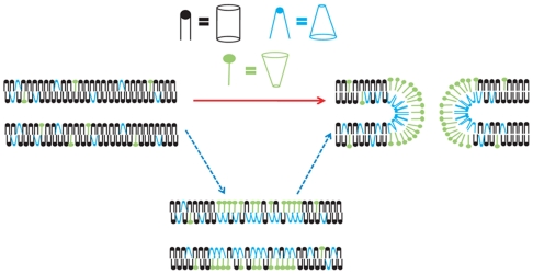
Mass spectrometry imaging has been used here to suggest that changes in membrane structure drive lipid domain formation in mating single-cell organisms. Chemical studies of lipid bilayers in both living and model systems have revealed that chemical composition is coupled to localized membrane structure. However, it is not clear if the lipids that compose the membrane actively modify membrane structure or if structural changes cause heterogeneity in the surface chemistry of the lipid bilayer. We report that time-of-flight secondary ion mass spectrometry images of mating Tetrahymena thermophila acquired at various stages during mating demonstrate that lipid domain formation, identified as a decrease in the lamellar lipid phosphatidylcholine, follows rather than precedes structural changes in the membrane. Domains are formed in response to structural changes that occur during cell-to-cell conjugation. This observation has wide implications in all membrane processes.
Conflict of interest statement
The authors declare no conflict of interest.
Figures

References
-
- Blumenthal R, Clague MJ, Durell SR, Epand RM. Membrane fusion. Chem Rev. 2003;103(1):53–69. - PubMed
-
- Yang L, Huang HW. Observation of a membrane fusion intermediate structure. Science. 2002;297(5588):1877–1879. - PubMed
-
- Simons K, Ikonen E. Functional rafts in cell membranes. Nature. 1997;387(6633):569–572. - PubMed
Publication types
MeSH terms
Substances
Grants and funding
LinkOut - more resources
Full Text Sources
Molecular Biology Databases
