. 2010 Feb 23;107(8):3441-6.
doi: 10.1073/pnas.0914798107. Epub 2010 Feb 1.

Bifurcation of insulin signaling pathway in rat liver: mTORC1 required for stimulation of lipogenesis, but not inhibition of gluconeogenesis

Affiliations

Bifurcation of insulin signaling pathway in rat liver: mTORC1 required for stimulation of lipogenesis, but not inhibition of gluconeogenesis

Shijie Li et al. Proc Natl Acad Sci U S A. .

Abstract

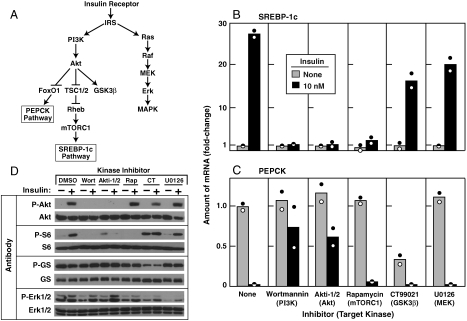
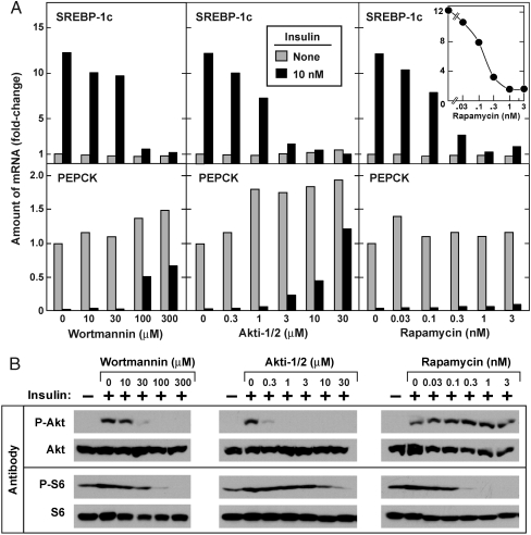
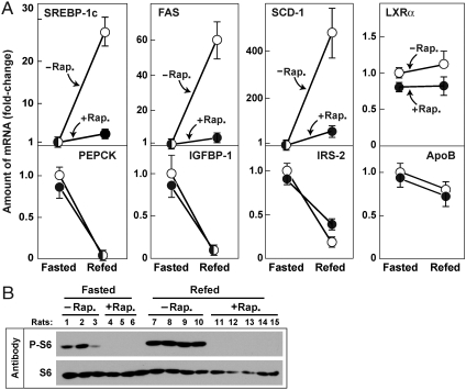
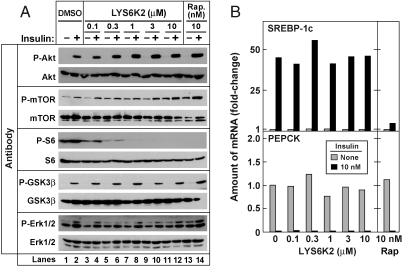
The livers of insulin-resistant, diabetic mice manifest selective insulin resistance, suggesting a bifurcation in the insulin signaling pathway: Insulin loses its ability to block glucose production (i.e., it fails to suppress PEPCK and other genes of gluconeogenesis), yet it retains its ability to stimulate fatty acid synthesis (i.e., continued enhancement of genes of lipogenesis). Enhanced lipogenesis is accompanied by an insulin-stimulated increase in the mRNA encoding SREBP-1c, a transcription factor that activates the entire lipogenic program. Here, we report a branch point in the insulin signaling pathway that may account for selective insulin resistance. Exposure of rat hepatocytes to insulin produced a 25-fold increase in SREBP-1c mRNA and a 95% decrease in PEPCK mRNA. Insulin-mediated changes in both mRNAs were blocked by inhibitors of PI3K and Akt, indicating that these kinases are required for both pathways. In contrast, subnanomolar concentrations of rapamycin, an inhibitor of the mTORC1 kinase, blocked insulin induction of SREBP-1c, but had no effect on insulin suppression of PEPCK. We observed a similar selective effect of rapamycin in livers of rats and mice that experienced an insulin surge in response to a fasting-refeeding protocol. A specific inhibitor of S6 kinase, a downstream target of mTORC1, did not block insulin induction of SREBP-1c, suggesting a downstream pathway distinct from S6 kinase. These results establish mTORC1 as an essential component in the insulin-regulated pathway for hepatic lipogenesis but not gluconeogenesis, and may help to resolve the paradox of selective insulin resistance in livers of diabetic rodents.


Conflict of interest statement

The authors declare no conflict of interest.

Figures

Fig. 1.
Fig. 1.
Insulin-mediated stimulation of SREBP-1c expression and inhibition of PEPCK expression in primary rat hepatocytes: effect of protein kinase inhibitors. (A) Insulin-activated protein kinase cascades as they relate to the SREBP-1c pathway (lipogenesis) and the PEPCK pathway (gluconeogenesis). (BD) Hepatocytes were prepared and plated on day zero as described in Methods. On day one, cells were pretreated for 30 min with the following concentration of the indicated inhibitor: 0.1 μM wortmannin, 10 μM Akti-1/2, 0.1 μM rapamycin, 2 μM CT99021, or 10 μM U0126. Each well of cells then received insulin (final concentration, 10 nM), after which the cells were incubated for 6 h at 37 °C and then harvested. Duplicate wells of cells were pooled for measurement of mRNA by quantitative RT-PCR and phosphorylated proteins by immunoblot analysis. (B, C) Relative amounts of mRNAs for SREBP-1c (B) and PEPCK (C). Each value represents the amount of mRNA relative to that in cells receiving no inhibitor and no insulin. Each bar shows the average of two independent experiments (denoted by Circles) performed on different days. (D) Immunoblot analysis of phosphorylated Akt, S6 ribosomal protein (S6), glycogen synthase (GS), and Erk1/2. Filters were exposed to film for 2 sec (P-Akt, P-S6, and P-GS), 15 sec (Akt, S6, GS, and Erk1/2), or 30 sec (P-Erk1/2) at room temperature. Experiments in BD were done three times with similar results.
Fig. 2.
Fig. 2.
Dose-dependent effects of wortmannin, Akti-1/2, and rapamycin on SREBP-1c and PEPCK mRNA levels in primary rat hepatocytes. (A) Relative amounts of mRNAs for SREBP-1c (Top) and PEPCK (Bottom) in hepatocytes treated with the indicated concentration of the indicated inhibitor in the absence or presence of insulin as described in Fig. 1. (B) Immunoblot analysis of phosphorylated Akt and S6 ribosomal proteins. Whole-cell lysates were subjected to immunoblot analysis with the indicated antibody as described in Fig. 1D. Filters were exposed to film for 2 sec (P-Akt and P-S6) or 15 sec (Akt and S6). Experiments in A and B were done two times with similar results.
Fig. 3.
Fig. 3.
Effect of rapamycin on levels of mRNA encoding SREBP-1c and its target genes in livers of rats subjected to fasting and refeeding. Two groups of male Sprague–Dawley rats were fasted for 48 h. Six h prior to sacrifice, one of the groups received an intraperitoneal injection of 20 mg/kg rapamycin (•) and the other group received vehicle (○). One group continued fasting and the other group was refed with a high carbohydrate diet as described in Methods. After 6 h, all animals were anesthesized, and the livers were removed for measurement of mRNAs and phosphorylated proteins. (A) mRNA levels as determined by quantitative RT PCR. Each value represents the amount of mRNA relative to that of the vehicle-treated fasted group that is denoted arbitrarily as one. Values represent mean ± SEM of 3–5 rats in each group. (B) Immunoblot analysis of phosphorylated and total S6 ribosomal protein in livers of the rats used in (A). Filters were exposed to film for 2 sec (P-S6) or 15 sec (S6). Rap., rapamycin. A similar experiment was carried out in mice with similar results, and the experiment in rats was repeated once with similar results.
Fig. 4.
Fig. 4.
S6K inhibitor fails to block insulin-induced SREBP-1c mRNA expression in primary rat hepatocytes. (A) Immunoblot analysis of insulin-stimulated phosphorylation of the indicated protein in the presence of increasing concentrations of LYS6K2 (Lanes 1–12) or 10 nM rapamycin (Rap.) (Lanes 13, 14). Whole-cell lysates were subjected to immunoblot analysis with the indicated antibody. Filters were exposed to film for 2 sec (P-Akt and P-S6), 15 sec (Akt, mTOR, S6, GSK3ß, and Erk1/2), 30 sec (P-Erk1/2), or 60 sec (P-mTOR and P-GSK3ß). (B) Relative amounts of mRNAs of SREBP-1c (Top) and PEPCK (Bottom) in hepatocytes treated with the indicated concentration of LYS6K2 or 10 nM rapamycin in the absence or presence of insulin as described in Fig. 1. Data are expressed as described in Fig. 1B and C. This experiment was carried out in parallel with that in (A). Experiments in A and B were done three times with similar results.

Comment in

Similar articles

Cited by

References

    1. Reaven GM. Why syndrome X? From Harold Himsworth to the insulin resistance syndrome. Cell Metab. 2005;1:9–14. - PubMed
    1. Kahn BB, Flier JS. Obesity and insulin resistance. J Clin Invest. 2000;106:473–481. - PMC - PubMed
    1. Brown MS, Goldstein JL. Selective vs total insulin resistance: A pathogenic paradox. Cell Metab. 2008;7:95–96. - PubMed
    1. Shimomura I, et al. Decreased IRS-2 and increased SREBP-1c lead to mixed insulin resistance and sensitivity in livers of lipodystrophic and ob/ob mice. Mol Cell. 2000;6:77–86. - PubMed
    1. Matsumoto M, Han S, Kitamura T, Accili D. Dual role of transcription factor FoxO1 in controlling hepatic insulin sensitivity and lipid metabolism. J Clin Invest. 2006;116:2464–2472. - PMC - PubMed

Publication types

MeSH terms