Comparative Study
. 2010 Feb 16;107(7):2884-9.
doi: 10.1073/pnas.0912718107. Epub 2010 Feb 1.

No difference in kinetics of tau or histone phosphorylation by CDK5/p25 versus CDK5/p35 in vitro

Affiliations
Comparative Study

No difference in kinetics of tau or histone phosphorylation by CDK5/p25 versus CDK5/p35 in vitro

Dylan W Peterson et al. Proc Natl Acad Sci U S A. .

Abstract

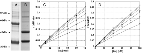
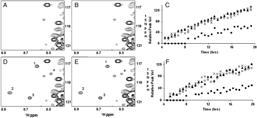
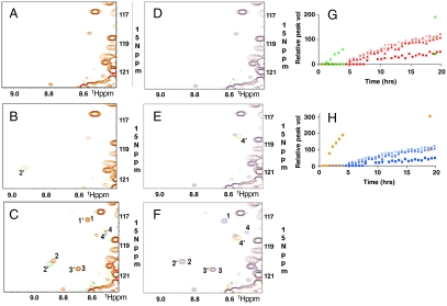
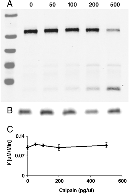
CDK5/p35 is a cyclin-dependent kinase essential for normal neuron function. Proteolysis of the p35 subunit in vivo results in CDK5/p25 that causes neurotoxicity associated with a number of neurodegenerative diseases. Whereas the mechanism by which conversion of p35 to p25 leads to toxicity is unknown, there is common belief that CDK5/p25 is catalytically hyperactive compared to CDK5/p35. Here, we have compared the steady-state kinetic parameters of CDK5/p35 and CDK5/p25 towards both histone H1, the best known substrate for both enzymes, and the microtubule-associated protein, tau, a physiological substrate whose in vivo phosphorylation is relevant to Alzheimer's disease. We show that the kinetics of both enzymes are the same towards either substrate in vitro. Furthermore, both enzymes display virtually identical kinetics towards individual phosphorylation sites in tau monitored by NMR. We conclude that conversion of p35 to p25 does not alter the catalytic efficiency of the CDK5 catalytic subunit by using histone H1 or tau as substrates, and that neurotoxicity associated with CDK5/p25 is unlikely attributable to CDK5 hyperactivation, as measured in vitro.


Conflict of interest statement

The authors declare no conflict of interest.

Figures

Fig. 1.
Fig. 1.
Steady-state kinetic analysis of tau phosphorylation by CDK5 bound to p35 or p25. A, B) SDS-PAGE analysis of baculovirus expressed His6CDK5/GSTp35 (A) and His6CDK5/GSTp25 (B). Enzymes were purchased from Millipore. (C, D) Kinetic analysis of tau phosphorylation by His6CDK5/GSTp35 (8.5 nM) (C) and His6CDK5/GSTp25 (12 nM) (D). Initial rates were obtained in response to varying both tau and ATP concentrations. Data were analyzed by global nonlinear regression fitting to give the best-fit parameters in Table 2. In both C and D, ATP concentrations are (from top to bottom): 1 mM -x, 500 -♦, 250 -□, 125 -▴, 62.5 -○, and 31 uM -▾
Fig. 2.
Fig. 2.
1H-15N-HSQC spectra of tau phosphorylated with CDK5/p35 or CDK5/p25. Region of new crosspeaks in response to phosphorylation by CDK5/p35 or CDK5/p25 is shown: A, D) CDK5/p35 (11 nM); B, E) CDK5/p25 (11 nM); A, B) after 30 min phosphorylation; D, E) after 19 hrs phosphorylation. New crosspeaks are labeled 1–4; C, F) Time courses of phosphorylation monitored by increase in volume of crosspeaks 1–4. 1 -○, 2 -▴, 3 -x, and 4 -▪. C) CDK5/p35 F) CDK5/p25. The full NMR spectrum is shown in Fig. S2.
Fig. 3.
Fig. 3.
CDK5/p25 phosphorylation-site optimization in tau. AC) S235 The sequence S235PSS was optimized for phosphorylation by CDK5/p25 by changing to S235PKK. 1H-15N-HSQC spectra of S235-optimized (Green) and wild type (Red) tau is shown after A) 0, B) 2, and C) 19 hrs phosphorylation by CDK5/p25 (11 nM). G) Relative change in peak volumes over time. Red—wild type; Green—mutant. DF) S202 The sequence S202PGT was optimized for phosphorylation by CDK5/p25 by changing to S202PKK. 1H-15N-HSQC spectra of S202-optimized (Orange) and wild-type (Blue) tau is shown after D) 0, E) 2, and F) 19 hrs phosphorylation by CDK5/p25 (11 nM). T205 is deleted; therefore peak 1′ is not seen. H) Relative change in peak volumes over time. Blue—wild-type; Orange—mutant. Peaks 1, 2, 3, and 4—wild-type tau. Peaks 1′, 2′, 3′, and 4′—catalytically optimized tau. 1,1′,—pT205; 2,2′,—pS235; 3,3′,—pS404; and 4,4′,—pS202.
Fig. 4.
Fig. 4.
Enzymatic activity of CDK5 upon conversion of p35 to p25 by calpain. Aliquots of CDK5/p35 were incubated with varying amounts of calpain, the reaction stopped with ALLN, and then assayed for tau kinase activity. A) Western blot of p35. Markers are 95, 72, 55, 43, and 34 kDa (top-bottom). B) Western blot of CDK5. C) Initial velocity of CDK5 (10 nM) after varied degrees of cleavage of p35 to p25. Cleavage of p35 to p25 results in no change in CDK5 catalytic efficiency.

Similar articles

Cited by

References

    1. Ishiguro K, et al. A serine/threonine proline kinase activity is included in the tau protein kinase fraction forming a paired helical filament epitope. Neurosci Lett. 1991;128:195–8. - PubMed
    1. Ishiguro K, et al. Tau protein kinase I converts normal tau protein into A68-like component of paired helical filaments. J Biol Chem. 1992;267:10897–901. - PubMed
    1. Lew J, Beaudette K, Litwin CM, Wang JH. Purification and characterization of a novel proline-directed protein kinase from bovine brain. J Biol Chem. 1992;267:13383–90. - PubMed
    1. Lew J, Winkfein RJ, Paudel HK, Wang JH. Brain proline-directed protein kinase is a neurofilament kinase which displays high sequence homology to p34cdc2. J Biol Chem. 1992;267:25922–6. - PubMed
    1. Lew J, Wang JH. Neuronal cdc2-like kinase. Trends Biochem Sci. 1995;20:33–7. - PubMed

Publication types