. 2010 Feb 16;107(7):2860-5.
doi: 10.1073/pnas.0915021107. Epub 2010 Feb 1.

Structure of Vibrio cholerae ToxT reveals a mechanism for fatty acid regulation of virulence genes

Affiliations

Structure of Vibrio cholerae ToxT reveals a mechanism for fatty acid regulation of virulence genes

Michael J Lowden et al. Proc Natl Acad Sci U S A. .

Abstract

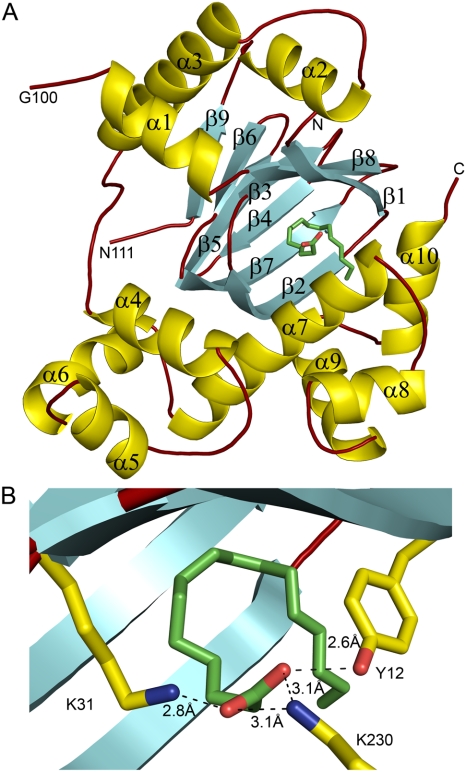
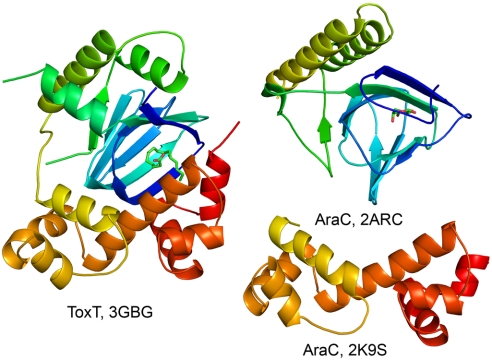
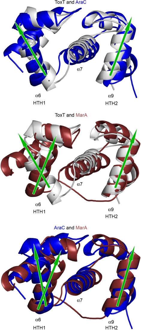
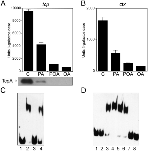
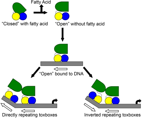
Cholera is an acute intestinal infection caused by the bacterium Vibrio cholerae. In order for V. cholerae to cause disease, it must produce two virulence factors, the toxin-coregulated pilus (TCP) and cholera toxin (CT), whose expression is controlled by a transcriptional cascade culminating with the expression of the AraC-family regulator, ToxT. We have solved the 1.9 A resolution crystal structure of ToxT, which reveals folds in the N- and C-terminal domains that share a number of features in common with AraC, MarA, and Rob as well as the unexpected presence of a buried 16-carbon fatty acid, cis-palmitoleate. The finding that cis-palmitoleic acid reduces TCP and CT expression in V. cholerae and prevents ToxT from binding to DNA in vitro provides a direct link between the host environment of V. cholerae and regulation of virulence gene expression.


Conflict of interest statement

The authors declare no conflict of interest.

Figures

Fig. 1.
Fig. 1.
The structure of ToxT. (A) Ribbon diagram of ToxT showing α-helices (gold), β-strands (cyan), and loops (dark red). The bound cis-palmitoleate is shown in stick form with carbons in green and oxygens in red. The N and C termini are indicated. Helices and strands are numbered according to their topological connectivity in the full-length protein. Note that residues 101–110 are disordered in the structure, as indicated by the loop ends on the left side of the molecule. (B) Close up of the cis-palmitoleate binding region showing interactions of the carboxylate head group with side chains.
Fig. 2.
Fig. 2.
Ribbon diagrams of ToxT and the N- and C-terminal domains of AraC. The models are colored by a rainbow effect with blue at the N terminus and red at the C terminus. The arabinose bound in the N-terminal domain of AraC is shown in stick form. PDB accession numbers are indicated for the three structures.
Fig. 3.
Fig. 3.
Pairwise SSM alignments of the DBDs of ToxT (3GBG), AraC (2K9S), and MarA (1BL0). ToxT is represented in silver, AraC in blue, and MarA in dark red. Helix α7 and the DNA-binding helices α6 and α9 are labeled. Arrows indicate the relative direction each DNA-binding helix is pointing. The asterisk indicates the distorted section of helix α6 on ToxT.
Fig. 4.
Fig. 4.
Effects of fatty acids on tcp and ctx expression. (A) and (B) β-galactosidase activity of tcp-lacZ and ctx-lacZ fusion constructs respectively. Cells were grown in LB pH 6.5 at 30 °C for 18 hours +/- the indicated fatty acids at 0.02% dissolved in methanol. The inset shows TcpA expression by Western blot under the same conditions in the corresponding lanes (C—control with methanol; PA—sodium palmitate; POA—palmitoleic acid; OA—oleic acid). (C) EMSA showing a specific interaction between ToxT and a 40-base-pair segment of the tcp promoter. All lanes contain 0.0025 μM labeled probe. Lane 1, free DNA; lane 2, 0.2 μM ToxT; lane 3, 0.2 μM ToxT with a 70-fold molar excess of cold competing DNA; lane 4, 0.2 μM ToxT with a 70-fold molar excess of a 42-base-pair nonspecific DNA cold competitor. (D) EMSA showing the effect of several fatty acids on ToxT/DNA interactions. All lanes contain 0.0025 μM labeled probe. Lane 1, free DNA; lane 2, 0.125 μM ToxT; lane 3, 0.2 μM ToxT; lane 4, 0.25 μM ToxT; lane 5, 0.25 μM ToxT with methanol; lane 6, 0.25 μM ToxT with 0.002% palmitic acid; lane 7, 0.25 μM ToxT with 0.002% palmitoleic acid; lane 8, 0.25 μM ToxT with 0.002% oleic acid.
Fig. 5.
Fig. 5.
Model for the regulation of ToxT by monounsaturated fatty acids. Fatty acid bound ToxT is in a “closed” conformation, which cannot bind to DNA. Release of the fatty acid results in an “open” conformation that can bind to DNA. In the “open” conformation the N-terminal domain is free to move in relation to the C-terminal domain, and is able to dimerize with another ToxT at an adjacent toxbox. The linker is sufficiently flexible to allow dimerization on DNA in either direct or inverted orientations.

Similar articles

Cited by

References

    1. Champion GA, Neely MN, Brennan MA, DiRita VJ. A branch in the ToxR regulatory cascade of Vibrio cholerae revealed by characterization of toxT mutant strains. Mol Microbiol. 1997;23:323–331. - PubMed
    1. DiRita VJ, Parsot C, Jander G, Mekalanos JJ. Regulatory cascade controls virulence in Vibrio cholerae. Proc Natl Acad Sci USA. 1991;88:5403–5407. - PMC - PubMed
    1. Higgins DE, Nazareno E, DiRita VJ. The virulence gene activator ToxT from Vibrio cholerae is a member of the AraC family of transcriptional activators. J Bacteriol. 1992;174:6974–6980. - PMC - PubMed
    1. Brown RC, Taylor RK. Organization of tcp, acf, and toxT genes within a ToxT-dependent operon. Mol Microbiol. 1995;16:425–439. - PubMed
    1. Yu RR, DiRita VJ. Analysis of an autoregulatory loop controlling ToxT, cholera toxin, and toxin-coregulated pilus production in Vibrio cholerae. J Bacteriol. 1999;181:2584–2592. - PMC - PubMed

Publication types

MeSH terms

LinkOut - more resources