. 2010 Mar 2;107(9):4099-104.
doi: 10.1073/pnas.0911904107. Epub 2010 Feb 1.

The transition state for folding of an outer membrane protein

Affiliations

The transition state for folding of an outer membrane protein

Gerard H M Huysmans et al. Proc Natl Acad Sci U S A. .

Abstract

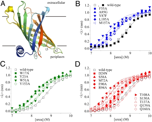
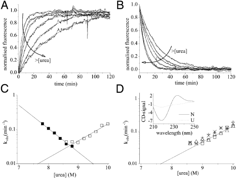
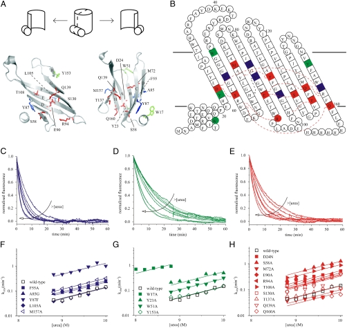
Inspired by the seminal work of Anfinsen, investigations of the folding of small water-soluble proteins have culminated in detailed insights into how these molecules attain and stabilize their native folds. In contrast, despite their overwhelming importance in biology, progress in understanding the folding and stability of membrane proteins remains relatively limited. Here we use mutational analysis to describe the transition state involved in the reversible folding of the beta-barrel membrane protein PhoPQ-activated gene P (PagP) from a highly disordered state in 10 M urea to a native protein embedded in a lipid bilayer. Analysis of the equilibrium stability and unfolding kinetics of 19 variants that span all eight beta-strands of this 163-residue protein revealed that the transition-state structure is a highly polarized, partly formed beta-barrel. The results provide unique and detailed insights into the transition-state structure for beta-barrel membrane protein folding into a lipid bilayer and are consistent with a model for outer membrane protein folding via a tilted insertion mechanism.


Conflict of interest statement

The authors declare no conflict of interest.

Figures

Fig. 1.
Fig. 1.
(A) Cartoon representation of PagP [Protein Data Bank (PDB) ID code 1THQ (19); W. L. DeLano, http://www.pymol.org (2002)]; β-strands are labeled A–H; periplasmic turns t1–3. (B) Equilibrium refolding (▪) and unfolding (□) of wild-type PagP. The unfolding transitions of PagP variants bearing mutations in residues in (B) the hydrophobic surface, (C) the aromatic girdles, and (D) the barrel interior are also shown. For these variants residues mutated that are located in the N-terminal half of the protein sequence are indicated by closed symbols, whereas residues in the C-terminal half of the protein sequence are shown as open symbols. Solid lines are global fits to a two-state mechanism yielding a common MUN = 6.86 ± 0.20 kJ/mol/M. All experiments were performed using 0.4 μM PagP in diC12∶0PC-liposomes at an LPR of 3200∶1 in 50 mM sodium phosphate buffer pH 8 at 25 °C.
Fig. 2.
Fig. 2.
(A) Folding and (B) unfolding kinetics of 0.4 μM PagP in diC12∶0PC-liposomes (LPR 3200∶1) measured by Trp-fluorescence. The arrows indicate increasing urea concentrations (7.8 to 8.8 M and 9 to 10 M urea in steps of 0.2 M for folding and unfolding, respectively). (C) Urea-concentration dependence of the rate constants of folding (▪) and unfolding (□). Lines represent linear fits to each dataset. (D) Urea-concentration dependence of the unfolding rate constant of PagP from diC12∶0PC-liposomes measured using Trp-fluorescence [0.4 μM PagP; LPR 3200∶1 (□) or 800∶1 (Δ)] and using CD-spectroscopy (5 μM PagP; LPR 800∶1) at 232 nm (×) and 218 nm (+). Inset, CD-spectra of native (N) and unfolded, membrane-associated (U) PagP [CD-signal = mean residue[Θ] × 103 (deg cm2 dmol-1)].
Fig. 3.
Fig. 3.
(A) Cartoon representation of a cross-section through PagP [PDB ID code 1THQ (19); W. L. DeLano, http://www.pymol.org (2002)]. A schematic is shown above to allow orientation. Mutated residues on the hydrophobic surface are highlighted in blue, those in the aromatic girdles are shown in green, and residues in the β-barrel interior are in red. (B) Topology map of PagP highlighting mutated residues colored as in (A) with their specific interactions (hydrogen or π-cation bonds) with other side chains (red dashed lines; see Fig. S3 for details). (CE) Examples of unfolding transients between 8.8 and 10 M urea (0.4 μM PagP; LPR 3200∶1) of F55A, Y153A, and R94A, respectively. The arrows indicate increasing urea concentrations in 0.2 M steps. (FH) Urea-concentration dependence of the unfolding rate constants of wild-type PagP and PagP variants bearing mutations in the hydrophobic exterior of the barrel, the aromatic girdles, and the barrel interior, respectively. Residues located in the N-terminal half of the β-barrel are indicated by closed symbols, residues in the C-terminal half by open symbols. The data were globally fitted yielding a common mu = 2.52 ± 0.11 kJ·mol-1·M-1.
Fig. 4.
Fig. 4.
(A) Cartoon representation of PagP [PDB ID code 1THQ (19); W. L. DeLano, http://www.pymol.org (2002)] with ΦF-values mapped onto the native structure. (B) Topology map of PagP highlighting the ΦF-values of mutated residues. (C) Proposed folding mechanism of PagP into membranes: unfolded, but membrane-associated PagP (I); tilted insertion of the transition-state ensemble (II); and assembly of the helical clamp to yield native PagP (III). Note that the periplasmic halves of strands A, B, C, and D are depicted as loops to depict the low ΦF-values of side-chains in this region of the transition-state structure. In AC, ΦF > 0.5 are in blue, 0.3 ≤ ΦF ≤ 0.5 are in purple, ΦF < 0.3 are in red, and ΦF < 0 are in orange. Residues 51, 72, 108, 137, and 160 gave formula image 
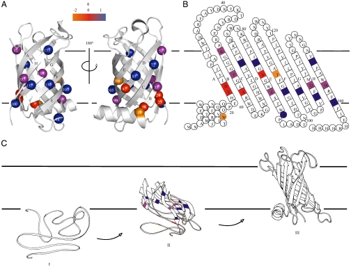
[the cut-off generally accepted to yield highly reliable ΦF-values (41)].

Comment in

  • Membrane protein folding makes the transition.
    Booth PJ, Clarke J. Booth PJ, et al. Proc Natl Acad Sci U S A. 2010 Mar 2;107(9):3947-8. doi: 10.1073/pnas.0914478107. Epub 2010 Feb 19. Proc Natl Acad Sci U S A. 2010. PMID: 20173094 Free PMC article. No abstract available.

References

    1. Wallin E, von Heijne G. Genome-wide analysis of integral membrane proteins from eubacterial, archaean, and eukaryotic organisms. Protein Sci. 1998;7:1029–1038. - PMC - PubMed
    1. Hediger MA, et al. The ABCs of solute carriers: Physiological, pathological and therapeutic implications of human membrane transport proteins. Pflugers Arch. 2004;447:465–468. - PubMed
    1. Sanders C, Myers JK. Disease-related misassembly of membrane proteins. Annu Rev Bioph Biom. 2004;33:25–51. - PubMed
    1. Chen GQ, Gouaux E. Probing the folding and unfolding of wild-type and mutant forms of bacteriorhodopsin in micellar solutions: Evaluation of reversible unfolding conditions. Biochemistry. 1999;38:15380–15387. - PubMed
    1. Bowie JU. Solving the membrane protein folding problem. Nature. 2005;438:581–589. - PubMed

Publication types

Substances