. 2010 Feb 23;107(8):3293-8.
doi: 10.1073/pnas.0906501107. Epub 2010 Feb 1.

Supramolecular design of self-assembling nanofibers for cartilage regeneration

Affiliations

Supramolecular design of self-assembling nanofibers for cartilage regeneration

Ramille N Shah et al. Proc Natl Acad Sci U S A. .

Abstract

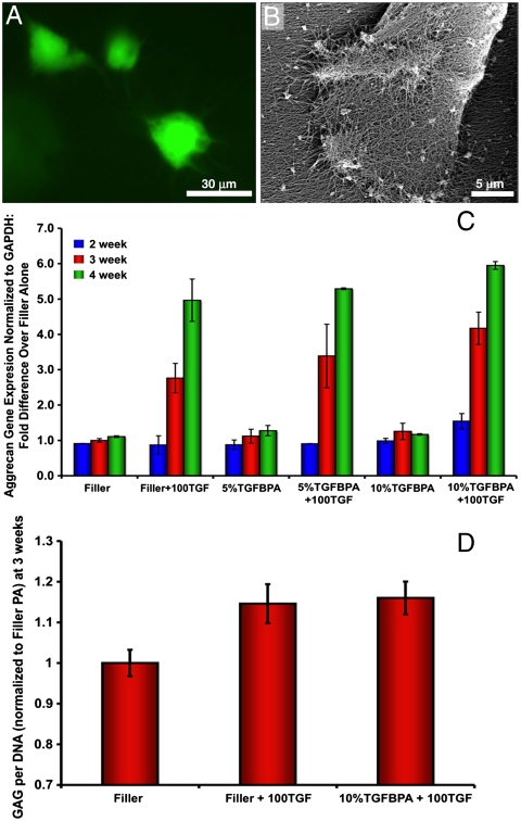
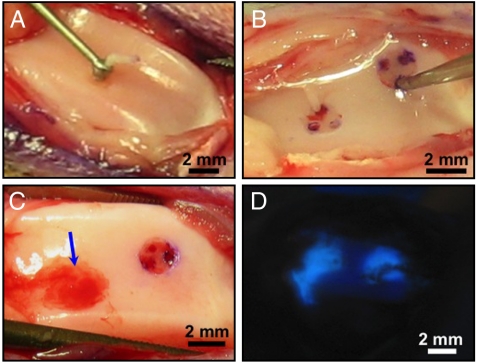
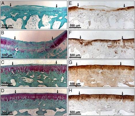
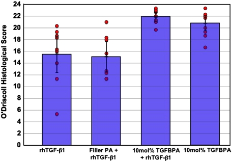
Molecular and supramolecular design of bioactive biomaterials could have a significant impact on regenerative medicine. Ideal regenerative therapies should be minimally invasive, and thus the notion of self-assembling biomaterials programmed to transform from injectable liquids to solid bioactive structures in tissue is highly attractive for clinical translation. We report here on a coassembly system of peptide amphiphile (PA) molecules designed to form nanofibers for cartilage regeneration by displaying a high density of binding epitopes to transforming growth factor beta-1 (TGFbeta-1). Growth factor release studies showed that passive release of TGFbeta-1 was slower from PA gels containing the growth factor binding sites. In vitro experiments indicate these materials support the survival and promote the chondrogenic differentiation of human mesenchymal stem cells. We also show that these materials can promote regeneration of articular cartilage in a full thickness chondral defect treated with microfracture in a rabbit model with or even without the addition of exogenous growth factor. These results demonstrate the potential of a completely synthetic bioactive biomaterial as a therapy to promote cartilage regeneration.


Conflict of interest statement

The authors declare no conflict of interest.

Figures

Fig. 1.
Fig. 1.
Design of PAs for articular cartilage regeneration. Chemical structure of (A) TGF-binding PA and (B) nonbioactive filler PA. (C) Illustration of coassembly of the TGF-binding PA and the filler PA showing binding epitopes exposed on the surface of the nanofiber. (D) ELISA results showing TGFβ-1 release up to 72 hours from filler PA and 10 mol% TGF-binding PA (TGFBPA). 100 ng/mL of TGFβ-1 were loaded in all gels. Error bars equal standard deviation, n = 4.
Fig. 2.
Fig. 2.
In vitro viability and differentiation of hMSCs cultured in PA scaffolds. (A) Live/dead images of cells cultured in PA gels (green = live; red = dead). (B) SEM of hMSC on nanofiber gel surface. (C) Aggrecan gene expression from hMSCs cultures in PA gels at 2, 3, and 4 wks. Filler PA = filler; 100 ng/mL of TGF = 100TGF; 5 mol% TGFBPA = 5%TGFBPA; 10 mol% TGFBPA = 10%TGFBPA. (D) GAG per DNA quantification in digested PA gels at 3 wks for filler, filler + 100TGF, and 10%TGFBPA + 100TGF groups. Error bars equal standard deviation, n = 3.
Fig. 3.
Fig. 3.
Full thickness articular cartilage defect microfracture rabbit model. Surgical procedure creating (A) full thickness articular cartilage defects in rabbit trochlea using a microcurette and (B) microfracture holes through the subchondral bone using a microawl to induce bleeding into the defect. (C) PA gel in defect after injection (Arrow). (D) Pyrene-labeled PA gel illustrating containment of the gel within articular cartilage defects after injection.
Fig. 4.
Fig. 4.
Observation of articular cartilage defects 12 wks after treatment. Macroscopic views of articular cartilage defects after 12 wks postop treated with (A) 100 ng/mL TGF-β1 (100TGF), (B) filler PA + 100TGF, (C) 10%TGFBPA + 100TGF, and (D) 10%TGFBPA alone.
Fig. 5.
Fig. 5.
Histological evaluation of sample sections 12 wks after treatment. Safranin-O staining for glycosaminoglycans (AD) and type II collagen staining (EH) in articular cartilage defects treated with (A, E) 100 ng/mL TGF-β1 (100TGF), (B, F) filler PA + 100TGF, (C, G) 10%TGFBPA + 100TGF, and (D, H) 10%TGFBPA alone 12 wks postop.
Fig. 6.
Fig. 6.
Histological scores of 12 wk in vivo samples showing significantly higher scores for the groups treated with the 10% TGF-binding PA with or without growth factor. Circles represent scores for individual specimens in each group (n = 8–10). Of note is the narrow distribution of scores for the defect groups treated with the 10% TGFBPA compared to the wider spread in scores for those treated with 100 ng/mL rhTGF-β1 (100TGF) alone or filler PA + 100TGF.

Similar articles

Cited by

References

    1. Buckwalter JA, Mankin HJ. Articular cartilage: Degeneration and osteoarthritis, repair, regeneration, and transplantation. Instr Course Lect. 1998;47:487–504. - PubMed
    1. O’Driscoll SW. Current concepts review—The healing and regeneration of articular cartilage. J Bone Joint Surg Am. 1998;80:1795–1812. - PubMed
    1. Hunziker EB. Articular cartilage repair: Basic science and clinical progress. A review of the current status and prospects. Osteoarthr Cartilage. 2002;10:432–463. - PubMed
    1. Jackson DW, Simon TM, Aberman HM. Symptomatic articular cartilage degeneration: The impact in the new millennium. Clin Orthop Relat Res. 2001;391 (Suppl):S14–25. - PubMed
    1. Williams RJ, Harnly HW. Microfracture: Indications, technique, and results. Instr Course Lect. 2007;56:419–428. - PubMed

Publication types

LinkOut - more resources