Comparative Study
. 2010 Feb 16;107(7):3058-63.
doi: 10.1073/pnas.0812851107. Epub 2010 Jan 28.

Competing feedback loops shape IL-2 signaling between helper and regulatory T lymphocytes in cellular microenvironments

Affiliations
Comparative Study

Competing feedback loops shape IL-2 signaling between helper and regulatory T lymphocytes in cellular microenvironments

Dorothea Busse et al. Proc Natl Acad Sci U S A. .

Abstract

Cytokines are pleiotropic and readily diffusible messenger molecules, raising the question of how their action can be confined to specific target cells. The T cell cytokine interleukin-2 (IL-2) is essential for the homeostasis of regulatory T (Treg) cells that suppress (auto)immunity and stimulates immune responses mediated by conventional T cells. We combined mathematical modeling and experiments to dissect the dynamics of the IL-2 signaling network that links the prototypical IL-2 producers, conventional T helper (Th) cells, and Treg cells. We show how the IL-2-induced upregulation of high-affinity IL-2 receptors (IL-2R) establishes a positive feedback loop of IL-2 signaling. This feedback mediates a digital switch for the proliferation of Th cells and functions as an analog amplifier for the IL-2 uptake capacity of Treg cells. Unlike other positive feedbacks in cell signaling that augment signal propagation, the IL-2/IL-2R loop enhances the capture of the signal molecule and its degradation. Thus Treg and Th cells can compete for IL-2 and restrict its range of action through efficient cellular uptake. Depending on activation status and spatial localization of the cells, IL-2 may be consumed exclusively by Treg or Th cells, or be shared between them. In particular, a Treg cell can deprive a stimulated Th cell of its IL-2, but only when the cells are located in close proximity, within a few tens of micrometers. The present findings explain how IL-2 can play two distinct roles in immune regulation and point to a hitherto largely unexplored spatiotemporal complexity of cytokine signaling.


Conflict of interest statement

The authors declare no conflict of interest.

Figures

Fig. 1.
Fig. 1.
Reaction–diffusion model of IL-2 signaling. (A) Antigen-stimulated Th cells secrete IL-2 that can be captured by IL-2Rs constitutively expressed on Treg cells or induced on Th cells. (B) Processes governing the dynamics of extracellular IL-2 (I), unoccupied (R), and occupied (R) IL-2Rα.
Fig. 2.
Fig. 2.
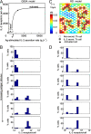
Digital IL-2Rα expression in Th cells. (A) The stimulus–response curve of IL-2Rα expression shows distinct basal and activated IL-2Rα levels (solid lines) that overlap in a small bi-stable region (dashed line, unstable state) (QSSA model). (B) Digital switching of IL-2Rα expression in a single cell translates into a binary response pattern of the cell population (QSSA model). For 1,000 cells the rates of antigen- and feedback-driven IL-2Rα expression were chosen from log-normal distributions (v10 = 150 ± 23 molecules/cell/h and v11 = 3,000 ± 762 molecules/cell/h) and increasing antigen stimulus modeled by q0 = 0, 300, 600, 1,800, 2,700, 15,000 molecules/cell/h (Top to Bottom). (C) Simulation of the RD model for 173 Th cells with 25% secreting IL-2 (q0 = 5,000 molecules/cell/h). The snapshot at 10 h after onset of stimulation shows complete autocrine and rare paracrine activation. (D) Digital distribution of IL-2Rα expression with, Top to Bottom, 10, 20, 30, 40, 50, and 60% IL-2 secreting cells (RD model).
Fig. 3.
Fig. 3.
Th cells exhibit digital IL-2Rα upregulation and proliferation. (A) Digital IL-2Rα expression pattern in Th-cell populations stimulated with increasing antigen doses. OVA-TCRtg/tg CD4+CD25 T cells were labeled with the proliferation marker CFSE and cultured with irradiated antigen-presenting cells and OVA peptide (concentration as indicated). Surface IL-2Rα was measured by flow cytometry at 72 h to correlate it with cell proliferation (1 representative experiment of 3). (B) Only cells with upregulated IL-2Rα proliferated, as seen by loss of CFSE intensity. Addition of blocking anti-IL-2 antibodies (αIL-2) to the cell culture prevented both IL-2Rα upregulation (C) and cell proliferation (D).
Fig. 4.
Fig. 4.
Interaction of Th and Treg cells through IL-2. (A) Bifurcation diagram computed for a proximal Th–Treg cell pair (QSSA model, L = 10 μm). The Th-cell activation threshold θ is increased and bi-stability enhanced (black line) (Fig. 2). By contrast, the upregulation of IL-2Rs on the Treg cell is practically continuous (red line). (B) Accordingly, bimodal expression of IL-2Rα on Th cells and gradual upregulation on Treg cells are predicted (parameter values as in Fig. 2). (C) Simulation of RD model for a Th–Treg coculture (cell ratio 2:1; 20% IL-2 producing Th cells, q0 = 5,000 molecules/cell/h; snapshot at 10 h after onset of stimulation). None of the Th cells become activated. (D) Upregulation of IL-2Rα on Th and Treg cells (RD model, fractions of IL-2-secreting Th cells were, Top to Bottom, 10, 20, 30, 40, 50, and 60%). Heterogeneity arises due to the random positioning of the different types of cell on a hexagonal array.
Fig. 5.
Fig. 5.
Distance dependence of paracrine IL-2 uptake by Treg cells. Behavior of a Th–Treg cell pair in dependence on IL-2 secretion rate by the Th cell and intercellular distance (Left) (QSSA model). The activation threshold for Th cells (θ, thick black line) and the line indicating strong IL-2 capture by Treg cells (thin black line, 10,000 upregulated IL-2Rα per cell) divide the phase diagram into four regions (see text).
Fig. 6.
Fig. 6.
IL-2 uptake by Treg cells. In a coculture of Th and Treg cells, IL-2Rα expression on Th cells is digital (A) whereas the expression of IL-2Rα in Treg cells adapts in a graded manner to the Th-cell stimulus (B). Aggrecan-TCRtg/tg CD4+CD25+ Treg cells were cocultured with CFSE-labeled OVA-TCRtg/tg CD4+CD25 Th cells at 1:2 ratio at different concentrations of OVA peptide (as indicated) and constant [aggrecan] = 2 μg/mL. Surface IL-2Rα was determined by flow cytometry (72 h) (1 representative experiment of 3). (C) Mean IL-2Rα expression and (D) fraction of active cells in the activated Th cells (blue lines) and Treg cells (red line) as determined by fitting the experimental IL-2Rα histograms. (E) Divided Th cells have elevated IL-2Rα. At 1 μg/mL OVA some activated Th cells already begin to lose IL-2Rα expression at 72 h so that some divided cells appear IL-2Rα-negative. The Treg cells have not been labeled with CFSE. The onset of IL-2Rα upregulation (F) and cell proliferation (G) are shifted to higher antigen stimuli by the presence of Treg cells, whereas a large stimulus (1 μg/mL OVA) overcomes the inhibitory Treg effect on the proliferation of the Th cells. Cell proliferation was quantified as ΔCFSE = MFI(generation 0) − MFI(generations >0); P-values for equal proliferation of Th cells and Treg cells are (t test): 0.017 (2.5 × 10⁻³ μg/mL OVA), 0.002 (0.01 μg/mL), 0.076 (0.05 μg/mL), and 0.359 (1 μg/mL).

References

    1. Cantrell DA, Smith KA. The interleukin-2 T-cell system: A new cell growth model. Science. 1984;224:1312–1316. - PubMed
    1. Malek TR. The biology of interleukin-2. Annu Rev Immunol. 2008;26:453–479. - PubMed
    1. Shevach EM. Regulatory T cells in autoimmunity. Annu Rev Immunol. 2000;18:423–449. - PubMed
    1. Sakaguchi S, Yamaguchi T, Nomura T, Ono M. Regulatory T cells and immune tolerance. Cell. 2008;133:775–787. - PubMed
    1. Lu LF, Rudensky A. Molecular orchestration of differentiation and function of regulatory T cells. Genes Dev. 2009;23:1270–1282. - PMC - PubMed

Publication types

Substances

LinkOut - more resources