. 2010 Feb 16;107(7):3186-91.
doi: 10.1073/pnas.0914876107. Epub 2010 Jan 26.

DJ-1 protects the nigrostriatal axis from the neurotoxin MPTP by modulation of the AKT pathway

Affiliations

DJ-1 protects the nigrostriatal axis from the neurotoxin MPTP by modulation of the AKT pathway

Hossein Aleyasin et al. Proc Natl Acad Sci U S A. .

Abstract

Loss-of-function DJ-1 (PARK7) mutations have been linked with a familial form of early onset Parkinson disease. Numerous studies have supported the role of DJ-1 in neuronal survival and function. Our initial studies using DJ-1-deficient neurons indicated that DJ-1 specifically protects the neurons against the damage induced by oxidative injury in multiple neuronal types and degenerative experimental paradigms, both in vitro and in vivo. However, the manner by which oxidative stress-induced death is ameliorated by DJ-1 is not completely clear. We now present data that show the involvement of DJ-1 in modulation of AKT, a major neuronal prosurvival pathway induced upon oxidative stress. We provide evidence that DJ-1 promotes AKT phosphorylation in response to oxidative stress induced by H(2)O(2) in vitro and in vivo following 1-methyl-4-phenyl-1,2,3,6-tetrahydropyridine (MPTP) treatment. Moreover, we show that DJ-1 is necessary for normal AKT-mediated protective effects, which can be bypassed by expression of a constitutively active form of AKT. Taken together, these data suggest that DJ-1 is crucial for full activation of AKT upon oxidative injury, which serves as one explanation for the protective effects of DJ-1.


Conflict of interest statement

The authors declare no conflict of interest.

Figures

Fig. 1.
Fig. 1.
AKT activation is suppressed in the absence of DJ-1. (A) Cortical neurons from DJ-1+/+ and DJ-1−/− embryos were harvested, plated, and treated with H2O2 (100 μM) in a time-dependent fashion. Extracts were probed for pAKT (S473), total AKT, and β-actin by Western blot. (B) Quantification of A from three independent experiments. Values are presented as mean optical density relative to total AKT. (C) Eight- to 10-week-old C57Bl6 mice of WT and DJ-1 knockout genotype were treated with two 30-mg/kg doses of MPTP (M), or saline (S), given 24 h apart. Three hours following the second injection, mice brains were quickly dissected for SNc and samples were processed for Western blot analysis. (D) Quantification of C. n = 3–6 per group. (E) Immortalized lymphoblasts derived from patients with the DJ-1 L166P mutation or healthy control lymphoblasts were treated with H2O2 in a time dependant manner. Analysis of cell lysates was carried out by Western blot. Blot presented is representative of two independent experiments. Data are presented as mean ± SEM.
Fig. 2.
Fig. 2.
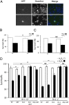
AKT requires DJ-1 to exert its neuroprotective function in vitro. (A) Representative pictures of alive (Upper, large arrowhead) and dead (Lower, thin arrowhead) neurons. Neuronal survival was measured by identifying GFP-positive cells and determining their nuclear integrity by Hoechst stain. (B–D) Cortical neurons from either DJ-1+/+ or DJ-1−/− embryos were harvested, plated, and transfected with empty vector (EV), AKT, DJ-1, or Myr AKT. Cells were treated with H2O2 (30 μM) or vehicle control (−H2O2) for 3 h. Quantification was assessed as in A. Data are presented as mean ± SEM. **, P < 0.01; NS, no significant difference.
Fig. 3.
Fig. 3.
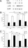
DJ-1 requires AKT activation to promote cellular survival in vitro. (A) Cortical neurons were treated for either 10 or 20 μM of LY with and without H2O2 (100 μM, 15 min) to determine the effective dose of LY. (B) Cortical neurons were infected with either GFP or DJ-1 with GFP. Cells were then pretreated with LY followed by H2O2 treatment for survival assessment. (C) Cortical neurons were cotransfected with GFP and empty vector (EV), DJ-1, DN-AKT, or a DJ-1/DN-AKT combination followed by H2O2 treatment. Survival was assessed as in B. Data are presented as mean ± SEM. *, P < 0.05; **, P < 0.01; NS, no significant difference.
Fig. 4.
Fig. 4.
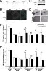
AKT requires DJ-1 to exert its neuroprotective function in an in vivo model of PD. (A) Schematic representation of treatment course. Mice were injected ipsilaterally in the striatum with adenovirus (LacZ, HA-AKT, Myr-AKT) 7 d before commencement of MPTP injections. MPTP was injected for 5 consecutive days and brains were collected 14 days following the first MPTP injection. (B) Confirmation of virus expression was performed by immunohistochemistry. Dual labeling of both TH and protein of interest in the SNc. (C) HA expression was tested in the SNc by Western blot analysis. (D) Representative pictures of both striatum and SNc of mice treated with MPTP or saline. SNc and striatum were stained for TH and DAT, respectively. (E) Quantification of TH-immunoreactive neurons was performed at the MTN region of the SNc where virus expression was highest. “Ipsi” denotes the side of the brain ipsilateral to the virus injection, whereas “contra” denotes the contralateral side. (F) Quantification of DAT-positive fibers normalized to cortex (DAT-negative). Data are presented as mean ± SEM. *, P < 0.05; **, P < 0.01; NS, no significant difference.
Fig. 5.
Fig. 5.
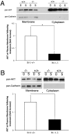
AKT requires DJ-1 to localize to membranous fractions following oxidative insult. (A) DJ-1+/+ and DJ-1−/− MEFs were treated with 500 μM of H2O2 for 5 min or with media control. Western blot analysis by probing pan-AKT and pan-Cadherin as membranous fraction control. Quantification of membranous fractions was performed in the lower panel by calculating relative AKT density normalized to Cadherin levels and normalizing treatment group to control. Data are representative of n = 4 experiments. (B) DJ-1+/+ and DJ-1−/− cortical neurons were subjected to 100 μM of H2O2 for 5 min or media control. Quantification was performed as in A. Data are presented as mean ± SEM. *, P < 0.05.

Similar articles

Cited by

References

    1. Bonifati V, et al. Mutations in the DJ-1 gene associated with autosomal recessive early-onset parkinsonism. Science. 2003;299:256–259. - PubMed
    1. Abou-Sleiman PM, Healy DG, Quinn N, Lees AJ, Wood NW. The role of pathogenic DJ-1 mutations in Parkinson’s disease. Ann Neurol. 2003;54:283–286. - PubMed
    1. Gu L, et al. Involvement of ERK1/2 signaling pathway in DJ-1-induced neuroprotection against oxidative stress. Biochem Biophys Res Commun. 2009;383:469–474. - PubMed
    1. Inden M, et al. PARK7 DJ-1 protects against degeneration of nigral dopaminergic neurons in Parkinson’s disease rat model. Neurobiol Dis. 2006;24:144–158. - PubMed
    1. Lev N, et al. DJ-1 protects against dopamine toxicity. J Neural Transm. 2009;116:151–160. - PubMed

Publication types