

Expression of cancer testis antigen CT45 in classical Hodgkin lymphoma and other B-cell lymphomas
- PMID: 20133697
- PMCID: PMC2840360
- DOI: 10.1073/pnas.0915050107
Expression of cancer testis antigen CT45 in classical Hodgkin lymphoma and other B-cell lymphomas
Abstract
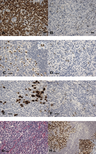
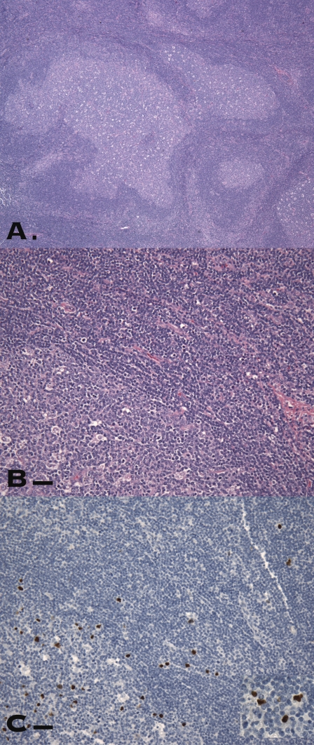
We have shown previously that cancer/testis (CT) antigen, CT45, is expressed in various epithelial cancers at a frequency of <5% to approximately 35%. In this study, the protein expression of CT45 was examined in non-Hodgkin B-cell lymphomas and classical Hodgkin lymphoma by immunohistochemical analysis. Serological response to CT45 was also evaluated by ELISA using CT45 recombinant protein and sera from patients with Hodgkin lymphoma. None of the 80 low-grade B-cell lymphomas, including chronic lymphocytic leukemia/small lymphocytic lymphoma, follicular lymphoma, and mantle cell lymphoma, expressed CT45. In comparison, CT45 was expressed in 28 of 126 (22%) diffuse large B-cell lymphomas (DLBCL). A remarkably high percentage (42/72, 58%) of classical Hodgkin lymphoma contained CT45-positive Reed-Sternberg cells. Nodular sclerosis and mixed-cellularity subtypes had similar frequency of CT45 expression, but most EBV-positive cases were CT45 negative. Gray-zone lymphoma (cases with features of both DLBCL and classical Hodgkin lymphoma) also showed frequent (64%) CT45 expression. Evaluation of reactive lymphoid tissues showed scattered CT45-positive lymphocytes in a single case of florid follicular hyperplasia, raising the possibility that this case was an evolving malignancy. Despite frequent CT45 expression, only 1 of 67 Hodgkin lymphoma patients had detectable anti-CT45 antibodies in the serum, suggesting that the immune response to CT45 may be suppressed. In conclusion, classical Hodgkin lymphoma has the highest frequency of CT45 expression among all malignancies tested to date, the frequency of CT45 expression in DLBCL is similar to that seen in epithelial cancers, and low-grade non-Hodgkin B-cell lymphomas do not express CT45.
Conflict of interest statement
The authors declare no conflict of interest.
Figures
Similar articles
-
Characterization and expression of CT45 in Hodgkin's lymphoma.Clin Cancer Res. 2006 Aug 15;12(16):4804-11. doi: 10.1158/1078-0432.CCR-06-0186. Clin Cancer Res. 2006. PMID: 16914565
-
Utility of CCR7 to differentiate classic Hodgkin lymphoma and other B-cell lymphomas by flow cytometry and immunohistochemistry.Am J Clin Pathol. 2025 Feb 12;163(2):266-276. doi: 10.1093/ajcp/aqae119. Am J Clin Pathol. 2025. PMID: 39288406
-
Connective tissue growth factor is expressed in malignant cells of Hodgkin lymphoma but not in other mature B-cell lymphomas.Am J Clin Pathol. 2010 Feb;133(2):271-80. doi: 10.1309/AJCPG7H0SSRYKNKH. Am J Clin Pathol. 2010. PMID: 20093237
-
Reed-Sternberg-Like Cells in Non-Hodgkin Lymphomas.Arch Pathol Lab Med. 2015 Oct;139(10):1205-10. doi: 10.5858/arpa.2015-0197-RAI. Arch Pathol Lab Med. 2015. PMID: 26414463 Review.
-
Complexities in the diagnosis of large B-cell lymphomas, classic Hodgkin lymphomas and overlapping peripheral T-cell lymphomas simplified: An evidence-based guide.Ann Diagn Pathol. 2020 Jun;46:151534. doi: 10.1016/j.anndiagpath.2020.151534. Epub 2020 May 15. Ann Diagn Pathol. 2020. PMID: 32473554
Cited by
-
DNA hypomethylation-mediated activation of Cancer/Testis Antigen 45 (CT45) genes is associated with disease progression and reduced survival in epithelial ovarian cancer.Epigenetics. 2015;10(8):736-48. doi: 10.1080/15592294.2015.1062206. Epigenetics. 2015. PMID: 26098711 Free PMC article.
-
Cancer/testis antigens expression and autologous serological response in a set of Brazilian non-Hodgkin's lymphoma patients.Cancer Immunol Immunother. 2012 Dec;61(12):2207-14. doi: 10.1007/s00262-012-1285-6. Epub 2012 May 26. Cancer Immunol Immunother. 2012. PMID: 22638551 Free PMC article.
-
Hodgkin's lymphoma RNA-transfected dendritic cells induce cancer/testis antigen-specific immune responses.Cancer Immunol Immunother. 2012 Oct;61(10):1769-79. doi: 10.1007/s00262-012-1239-z. Epub 2012 Mar 15. Cancer Immunol Immunother. 2012. PMID: 22419371 Free PMC article.
-
Cancer/testis (CT) antigens, carcinogenesis and spermatogenesis.Spermatogenesis. 2011 Jul-Sep;1(3):209-220. doi: 10.4161/spmg.1.3.17990. Epub 2011 Jul 1. Spermatogenesis. 2011. PMID: 22319669 Free PMC article. Review.
-
CT45A1 acts as a new proto-oncogene to trigger tumorigenesis and cancer metastasis.Cell Death Dis. 2014 Jun 5;5(6):e1285. doi: 10.1038/cddis.2014.244. Cell Death Dis. 2014. PMID: 24901056 Free PMC article.
References
-
- Boon T, Coulie PG, Van den Eynde B. Tumor antigens recognized by T cells. Immunol Today. 1997;18:267–268. - PubMed
-
- Simpson AJ, Caballero OL, Jungbluth A, Chen YT, Old LJ. Cancer/testis antigens, gametogenesis and cancer. Nat Rev Cancer. 2005;5:615–625. - PubMed
-
- van der Bruggen P, et al. A gene encoding an antigen recognized by cytolytic T lymphocytes on a human melanoma. Science. 1991;254:1643–1647. - PubMed
Publication types
MeSH terms
Substances
LinkOut - more resources
Full Text Sources
Medical
Molecular Biology Databases
