. 2010 Feb 16;107(7):3012-7.
doi: 10.1073/pnas.0914902107. Epub 2010 Jan 26.

Proliferative lesions and metalloproteinase activity in murine lupus nephritis mediated by type I interferons and macrophages

Affiliations

Proliferative lesions and metalloproteinase activity in murine lupus nephritis mediated by type I interferons and macrophages

Antigoni Triantafyllopoulou et al. Proc Natl Acad Sci U S A. .

Abstract

Glomerulonephritis is a major cause of morbidity in patients with systemic lupus erythematosus. Although substantial progress has been made in the identification of pathogenic triggers that result in autoantibody production, little is known about the pathogenesis of aggressive proliferative processes that lead directly to irreversible glomerular damage and compromise of renal function. In this study, we describe a model of polyinosinic: polycytidylic acid-accelerated lupus nephritis in NZB/W mice that is characterized by severe glomerular proliferative lesions with de novo crescent formation, findings that are linked with decreased survival and adverse outcomes in lupus. Proliferative glomerulonephritis was associated with infiltrating kidney macrophages and renal expression of IFN-inducible genes, matrix metalloproteinases (MMPs), and growth factors. Crescent formation and renal MMP and growth factor expression were dependent on renal macrophages that expressed Il10, MMPs, osteopontin, and growth factors, including Pdgfc and Hbegf. Infiltrating macrophages and renal MMP expression were induced by type I IFN. These findings reveal a role for type I IFNs and alternatively activated macrophages in aggressive proliferative lesions of lupus nephritis.


Conflict of interest statement

The authors declare no conflict of interest.

Figures

Fig. 1.
Fig. 1.
Poly (I:C) induces severe proliferative glomerulonephritis with crescents. Untreated nonproteinuric, untreated spontaneously proteinuric, and poly (I:C)-treated proteinuric NZB/W mice, were analyzed after 14 days of proteinuria. (A) Paraffin-embedded kidney sections stained for basement membrane and matrix constituents with periodic acid shiff stain (PAS). (B) Glomerular and interstitial histological scores. Each symbol indicates an individual mouse (n = 4–11 mice per group). **, P < 0.01. (C) Mice were treated with poly (I:C) for the indicated duration of treatment and analyzed after the indicated duration of proteinuria; kidney cortex mRNA was analyzed by real-time RT-PCR for ISG15 and STAT1; values are relative to expression of the gene encoding GAPDH (n = 4–6 mice per group). *, P < 0.05.
Fig. 2.
Fig. 2.
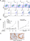
Poly (I:C) promotes kidney infiltration by CD11bhiGR1-F4/80+ macrophages. Untreated nonproteinuric, untreated spontaneously proteinuric and poly (I:C)-treated proteinuric NZB/W mice, were analyzed after 1, 4, and 14 days of proteinuria. (A) Whole-kidney single-cell suspensions were analyzed by FACS for expression of myeloid cell markers Gr1, CD11b, and F4/80. Representative dot plots from individual mice are shown. Percentages represent means ± SD (n = 3–6 mice per group). (B) Relative percentage of B cells, T cells, granulocytes, macrophages, and dendritic cells, per total kidney cells. Percentages represent means ± SD from three independent experiments (n = 3–9 mice per group). (C) Localization of CD11b+ cells in poly (I:C)-treated kidneys after 14 days of proteinuria. Representative of three independent experiments.
Fig. 3.
Fig. 3.
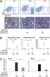
Macrophage depletion at the onset of proteinuria suppresses glomerular inflammation. Liposomal clodronate or PBS was injected i.v. at the onset of proteinuria (day 0) in poly (I:C)-treated mice. Injections were repeated after 3, 7, and 11 days of proteinuria. Untreated nonproteinuric, poly (I:C)-treated proteinuric, and poly (I:C) and liposomal clodronate treated proteinuric NZB/W mice, were analyzed after 14 days of proteinuria. (A) FACS analysis of whole kidney cells for expression of the myeloid/macrophage cell markers CD11b and GR1. Representative dot plots from individual mice are shown. Percentages represent means ± SD (n = 4–8 mice per group). Paraffin-embedded kidney sections, from the same kidneys analyzed by FACS, stained with PAS. (B) Glomerular and interstitial histological scores. Each symbol indicates an individual mouse. Results were pooled from three independent experiments (n = 10–12 mice per group). **, P < 0.01; ***, P < 0.001. (C) Kidney cortex mRNA was analyzed by real-time PCR for ISG15 and STAT1; values are relative to expression of the gene encoding GAPDH (n = 4–6 mice per group). *, P < 0.05.
Fig. 4.
Fig. 4.
Poly (I:C) promotes kidney metalloproteinase activity and up-regulates expression of metalloproteinases 2 and 14 in a macrophage-dependent manner. Liposomal clodronate or PBS was injected i.v. at the onset of proteinuria (day 0) in poly (I:C)- treated mice. Injections were repeated after 3, 7, and 11 days of proteinuria. Untreated nonproteinuric, poly (I:C)-treated proteinuric, and poly (I:C) and liposomal clodronate treated proteinuric NZB/W mice, were analyzed after 14 days of proteinuria. (A) Renal cortex mRNA was analyzed by real time PCR; values are relative to expression of the gene encoding GAPDH (n = 4–6 mice per group). *, P < 0.05; **, P < 0.01. (B) Gel zymography of nonreduced kidney cortex lysates. Each lane represents different individual mice. (C) Immunoblot analysis of MMP9, MMP2, and MMP14 in kidney cortex lysates. Tubulin was used as loading control. (D) In situ zymography with a gelatin substrate on unfixed kidney cortex cryosections. Representative of three independent experiments.
Fig. 5.
Fig. 5.
Poly (I:C) increases growth factor expression in a macrophage-dependent fashion. Kidney cortex mRNA was analyzed by real-time PCR for TGFβ, HB-EGF, PDGF-B, and osteopontin; values are relative to expression of the gene encoding GAPDH (n = 4–6 mice per group). *, P < 0.05; **, P < 0.01; ***, P < 0.001.
Fig. 6.
Fig. 6.
Poly (I:C)- induced kidney macrophages have a dysregulated tissue repair phenotype. (A) Whole kidney-cell suspensions were labeled with antibodies against lineage markers (B220, CD4, CD8, CD5, CD49b), as well as myeloid cell markers (CD11b, F4/80, CD11c) and analyzed by FACS. Lineage- (B220- CD4- CD8- CD5-CD49b-) CD11bhi CD11cintF4/80+ cells were sorted. (B) Total RNA from sorted kidney macrophages was analyzed by real-time PCR for IL10, TNFα, MMP2, MMP14, osteopontin, HB-EGF, and PDGF-C; values are relative to expression of the gene encoding GAPDH (n = 4–5 mice per group). *, P < 0.05.
Fig. 7.
Fig. 7.
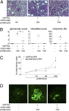
Type I IFN-mediated induction of proliferative crescentic nephritis. Untreated nonproteinuric, untreated spontaneously proteinuric and AdIFNα-treated proteinuric NZB/W mice were analyzed after 14 days of proteinuria. (A) Paraffin-embedded kidney sections stained for basement membrane and matrix constituents with PAS stain. (B) Glomerular and interstitial histologic scores. Each symbol indicates an individual mouse (n = 3–4 mice per group). *, P < 0.05. (C) Whole-kidney single-cell suspensions were analyzed by FACS after 1, 4, and 14 days of proteinuria. Relative percentage of B cells, T cells, granulocytes, macrophages, and dendritic cells per total kidney cells. Percentages are shown as the means ± SD from three independent experiments (n = 3–4 mice per group). (D) In situ zymography with a gelatin substrate on unfixed kidney cortex cryosections. Representative of three independent experiments.

Similar articles

Cited by

References

    1. Schwartz MM, Korbet SM, Katz RS, Lewis EJ. Evidence of concurrent immuno-pathological mechanisms determining the pathology of severe lupus nephritis. Lupus. 2009;18:149–158. - PubMed
    1. Kojo S, et al. Clinical usefulness of a prognostic score in histological analysis of renal biopsy in patients with lupus nephritis. J Rheumatol. 2009;36:2218–2223. - PubMed
    1. Hill GS, et al. Predictive power of the second renal biopsy in lupus nephritis: significance of macrophages. Kidney Int. 2001;59:304–316. - PubMed
    1. Baccala R, Hoebe K, Kono DH, Beutler B, Theofilopoulos AN. TLR-dependent and TLR-independent pathways of type I interferon induction in systemic autoimmunity. Nat Med. 2007;13:543–551. - PubMed
    1. Davidson A, Aranow C. Pathogenesis and treatment of systemic lupus erythematosus nephritis. Curr Opin Rheumatol. 2006;18:468–475. - PubMed

Publication types

MeSH terms

LinkOut - more resources