. 2010 Feb 23;107(8):3558-63.
doi: 10.1073/pnas.0910172106. Epub 2010 Feb 3.

Human and mouse adipose-derived cells support feeder-independent induction of pluripotent stem cells

Affiliations

Human and mouse adipose-derived cells support feeder-independent induction of pluripotent stem cells

Shigeki Sugii et al. Proc Natl Acad Sci U S A. .

Abstract

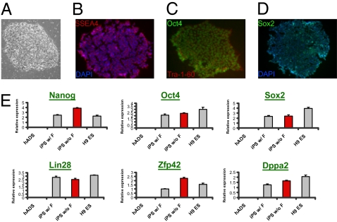
Although adipose tissue is an expandable and readily attainable source of proliferating, multipotent stem cells, its potential for use in regenerative medicine has not been extensively explored. Here we report that adult human and mouse adipose-derived stem cells can be reprogrammed to induced pluripotent stem (iPS) cells with substantially higher efficiencies than those reported for human and mouse fibroblasts. Unexpectedly, both human and mouse iPS cells can be obtained in feeder-free conditions. We discovered that adipose-derived stem cells intrinsically express high levels of pluripotency factors such as basic FGF, TGFbeta, fibronectin, and vitronectin and can serve as feeders for both autologous and heterologous pluripotent cells. These results demonstrate a great potential for adipose-derived cells in regenerative therapeutics and as a model for studying the molecular mechanisms of feeder-free iPS generation and maintenance.


Conflict of interest statement

The authors declare no conflict of interest.

Figures

Fig. 1.
Fig. 1.
Generation of ES-like iPS cell lines from mouse adipose-derived cells. (A) SVF Lin and Lin+ cells transduced with four reprogramming factors are stained for Nanog by immunohistochemistry after 7 days. Both whole-plate (Left) and two representative images of individual colonies (Right) are shown. Note that almost all Lin+-derived colonies are negative for Nanog. (B) mADS-derived iPS clones after derivation. (C) Gene expression analysis by qPCR indicates that two representative mADS-derived iPS clones express comparable pluripotent markers to those of mouse ES cells, in contrast to somatic cells, MEFs, mADS, and SVF cells.
Fig. 2.
Fig. 2.
In vivo pluripotency tests of mouse adipose-derived iPS cell lines. (AC) H&E staining of teratomas developed from mADS-derived iPS cells shows their contribution to ectoderm (B, brain; SE, squamous epithelium), mesoderm (Ad, adipose; Ct, cartilage; SM, smooth muscle), and endoderm (GC, goblet cells; P, pancreas). (D) A representative picture of a chimera mouse (right) produced from mADS-derived iPS cells. (E) Genotyping PCR analysis demonstrates the presence of retrovirus-specific DNA elements (LTR, long terminal repeat) in 50% of the offspring (nos. 2, 4, 6, 7, and 9) produced by crossing the chimera with wild-type mice. Endogenous GAPDH gene products are used as an internal control.
Fig. 3.
Fig. 3.
Reprogramming of human adipose-derived cells. (A) Development of hiPS lines from hADS cells (Upper) and hWP cells (Lower) after reprogramming factor introduction starting on day 0. (B) Gene expression analysis by qPCR shows that two independent hWP- and hADS-derived hiPS lines exhibit comparable levels of pluripotent markers to those in human keratinocyte-derived (hKerat) hiPS and H9 ES (hES) cells. (C) DNA methylation analysis of various Oct4 promoter regions indicates that somatic cells (hWP and hADS) are highly methylated whereas three independent hiPS (nos. 9, 10, and 12) clones derived from them are hypomethylated.
Fig. 4.
Fig. 4.
Characterization of human adipose-derived iPS cell lines produced in the feeder-free condition. (A) Morphology of a hADS-derived hiPS colony developed under a completely feeder-free condition. (BD) Representative immunofluorescence images of the feeder-free hADS-derived hiPS cells. Expression of ES cell surface antigens, SSEA4 and Tra-1-60 (B and C), and nuclear transcription factors, Oct4 and Sox2 (C and D), is observed. (E) qPCR analysis indicates that feeder-free hiPS cells from hADS cells (iPS w/o F) express pluripotent marker genes with levels comparable to those in hADS-hiPS cells made with feeder cells (iPS w/ F) or human ES cells (H9 ES).
Fig. 5.
Fig. 5.
Differentiation capacities of human adipose-derived iPS cells produced in the feeder-free condition. (AD) In vitro differentiation of feeder-free hiPS-derived embryoid bodies indicates markers of ectoderm, glial fibrillary acidic protein (GFAP) and beta III tubulin (Tuj) (A and B), of mesoderm, smooth muscle actin (SMA) (C), and of endoderm, alpha fetoprotein (AFP) (D). (EH) Feeder-free (E and F), hADS-derived (G), and hWP-derived (H) iPS cells exhibit in vivo differentiation capability and contribute to all three germ layers in teratomas. Ectoderm (B, brain; Ro, rosette; SE, squamous epithelium), mesoderm (Ct, cartilage; SM, smooth muscle; SkM, skeletal muscle), and endoderm (GC, goblet cells; P, pancreas) are shown.
Fig. 6.
Fig. 6.
High intrinsic pluripotency-supporting capabilities of adipose-derived stem cells. (A) Mouse adipose-derived stem (mADS) cells express high levels of self-renewal supporting factors that are comparable to those of MEFs by qPCR. (B) Human adipose-derived stem (hADS) cells express higher levels of self-renewal factors than most of other human cell lines. (C and D) hADS-derived iPS cells were grown for three passages on matrigel either in conditioned media (CM) taken from irradiated hADS cells, human foreskin fibroblasts (HFF), MEFs, or mADS cells (C) or by coculturing with these cells in Transwell plates (D). Flow cytometry using antibodies against SSEA3, SSEA4, and Tra-1-60 was performed to investigate relative expression of cell surface markers.

References

    1. Fraser JK, Wulur I, Alfonso Z, Hedrick MH. Fat tissue: An underappreciated source of stem cells for biotechnology. Trends Biotechnol. 2006;24:150–154. - PubMed
    1. Zuk PA, et al. Human adipose tissue is a source of multipotent stem cells. Mol Biol Cell. 2002;13:4279–4295. - PMC - PubMed
    1. Park IH, et al. Reprogramming of human somatic cells to pluripotency with defined factors. Nature. 2008;451:141–146. - PubMed
    1. Takahashi K, et al. Induction of pluripotent stem cells from adult human fibroblasts by defined factors. Cell. 2007;131:861–872. - PubMed
    1. Takahashi K, Yamanaka S. Induction of pluripotent stem cells from mouse embryonic and adult fibroblast cultures by defined factors. Cell. 2006;126:663–676. - PubMed

Publication types

LinkOut - more resources