. 2010 Feb 23;107(8):3487-92.
doi: 10.1073/pnas.0915166107. Epub 2010 Feb 3.

Identifying the amylome, proteins capable of forming amyloid-like fibrils

Affiliations

Identifying the amylome, proteins capable of forming amyloid-like fibrils

Lukasz Goldschmidt et al. Proc Natl Acad Sci U S A. .

Abstract

The amylome is the universe of proteins that are capable of forming amyloid-like fibrils. Here we investigate the factors that enable a protein to belong to the amylome. A major factor is the presence in the protein of a segment that can form a tightly complementary interface with an identical segment, which permits the formation of a steric zipper-two self-complementary beta sheets that form the spine of an amyloid fibril. Another factor is sufficient conformational freedom of the self-complementary segment to interact with other molecules. Using RNase A as a model system, we validate our fibrillogenic predictions by the 3D profile method based on the crystal structure of NNQQNY and demonstrate that a specific residue order is required for fiber formation. Our genome-wide analysis revealed that self-complementary segments are found in almost all proteins, yet not all proteins form amyloids. The implication is that chaperoning effects have evolved to constrain self-complementary segments from interaction with each other.


Conflict of interest statement

The authors declare no conflict of interest.

Figures
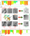
Fig. 1.
Fig. 1.
Validation of the 3D profile method for prediction of fibrillizing segments. The top and bottom panels show the predicted energy for fibrillation of every six-residue segment of RNase A. Red histogram bars represent hexapeptides with energy below -23 kcal/mol and are predicted to form fibrils. Seven such segments are shown, and each forms fibrils, as shown by the electron micrographs in the central panels. The black arrows indicate where three of these segments lie in the 3D structure of RNase A. This cartoon structure, like the histogram of energies, is colored with warmer colors representing a greater propensity for fibrillation. Blue and green segments are of higher energy and are predicted not to form fibrils. FERQHM is one of several segments predicted and experimentally confirmed not to form fibrils (first row of micrographs, right). However, when the residues of this segment were rearranged to QEMRHF (Table S2), the energy of the rearranged segment falls below -23 kcal/mol; QEMRHF is thus predicted to form fibrils, and it does (first row of micrographs). In addition, the fibril-forming segments QANKHI and STMSIT were rearranged to IHKAQN and ISMTTS, respectively, two sequences predicted and confirmed not to form fibrils (second and third row of micrographs). Notice that the longer SSTSAASSNY segment contains the SSTSAA segment, which forms fibrils and is capable of forming a steric zipper (Sawaya 2007). Taken together, the fibrillizing behavior of these 10 segments suggests that a threshold of -23 kcal/mol is appropriate for predicting fibrillation. The * indicates the C-terminal hinge loop, discussed in the text.
Fig. 2.
Fig. 2.
Sequence is more important than residue composition in determining propensity for formation of amyloid-like fibrils. (A) shows on the y axis the fibrillation propensity, calculated by the 3D profile method, and on the x axis the solvent exposure of segments in the nonredundant structures (proteins having less than 50% sequence identity) in the PDB. Segments below the horizontal -23 kcal/mol line are HP and are preferentially buried if < 15% exposed (vertical line). Thus buried HP segments are plotted in the lower left quadrant and are enclosed by a red rectangle. In (B) these segments are shuffled, and the fibrillation propensity and exposure of the resulting shuffled segments are plotted. The shuffling operation, which changes the residue order within a segment but does not alter its residue composition, allows these segments to migrate from the lower left quadrant to lower values of propensity (higher energy) and greater exposure to solvent. Only 42% of segments remain buried and retain their high fibrillogenic propensity. The exposure to solvent of these segments is estimated by examining the corresponding positions within proteins that contain the exact residue sequence. Of the 720 permutations for a given six-residue segment, about 12 are found in some PDB protein, and hence the solvent exposure can be determined and plotted. Warmer colors indicate higher count density. The percentage of segments found in each quadrant is indicated in white. The implications are that high propensity depends on a nonrandom pattern of sequences and that proteins tend to bury HP segments.

Similar articles

Cited by

References

    1. Astbury WT, Dickinson S, Bailey K. The x-ray interpretation of the denaturation and the structure of the seed globulihns. Biochem J. 1935;29:2351–2360. - PMC - PubMed
    1. Terry WD, et al. Structural identity of Bence Jones and amyloid fibril proteins in a patient with plasma cell dyscrasia and amyloidosis. J Clin Invest. 1973;52:1276–1281. - PMC - PubMed
    1. Sunde M, et al. Common core structure of amyloid fibrils by synchrotron x-ray diffraction. J Mol Biol. 1997;273:729–739. - PubMed
    1. Geddes AJ, Parker KD, Atkins ED, Beighton E. “Cross-beta” conformation in proteins. J Mol Biol. 1968;32:343–358. - PubMed
    1. Kelly J, Balch WE. Amyloid as a natural product. J Cell Biol. 2003;161:461–462. - PMC - PubMed

Publication types