. 2010 Feb 9;107(6):2550-5.
doi: 10.1073/pnas.0914508107. Epub 2010 Jan 20.

Molecular mimics can induce a nonautoaggressive repertoire that preempts induction of autoimmunity

Affiliations

Molecular mimics can induce a nonautoaggressive repertoire that preempts induction of autoimmunity

Emanual Maverakis et al. Proc Natl Acad Sci U S A. .

Abstract

To determine the role that competition plays in a molecular mimic's capacity to induce autoimmunity, we studied the ability of naïve encephalitogenic T cells to expand in response to agonist altered peptide ligands (APLs), some capable of stimulating both self-directed and exclusively APL-specific T cells. Our results show that although the APLs capable of stimulating exclusively APL-specific T cells are able to expand encephalitogenic T cells in vitro, the encephalitogenic repertoire is effectively outcompeted in vivo when the APL is used as the priming immunogen. Competition as a mechanism was supported by: (i) the demonstration of a population of exclusively APL-specific T cells, (ii) an experiment in which an encephalitogenic T cell population was successfully outcompeted by adoptively transferred naïve T cells, and (iii) demonstrating that the elimination of competing T cells bestowed an APL with the ability to expand naïve encephalitogenic T cells in vivo. In total, these experiments support the existence of a reasonably broad T cell repertoire responsive to a molecular mimic (e.g., a microbial agent), of which the exclusively mimic-specific component tends to focus the immune response on the invading pathogen, whereas the rare cross-reactive, potentially autoreactive T cells are often preempted from becoming involved.


Conflict of interest statement

The authors declare no conflict of interest.

Figures

Fig. 1.
Fig. 1.
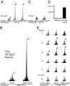
Characterization of the Ac1-9-specific Vβ8.2Jβ2.7 clonotype. (A) Mice were immunized with CFA alone or Ac1-9-CFA and draining lymph nodes were removed for analysis. Vβ8.2Jβ2.7 spectra are shown. Arrows indicate a TCR CDR3 length of 9 aa. Significant expansions within the Vβ8.2Jβ2.7 spectra were not seen when B10.PL animals were immunized with CFA alone. Likewise, when lymph node cells isolated from Ac1-9-CFA primed animals were incubated with medium alone, no significant expansions were found. In contrast, when lymph node cells from Ac1-9-CFA primed animals were cultured with Ac1-9, an expansion correlating to a CDR3 length of 9 aa was seen. This 9 aa expansion was also seen directly without the need of an in vitro culturing step when mononuclear cells were obtained from spinal cords isolated from mice suffering from EAE. This figure shows one of several similar experiments. (B) The MBP Ac1-9-specific Vβ8.2Jβ2.7 T cell hybridoma 172.10 can be stimulated by Ac1-6(M4), Ac1-9, and LDVM1-9(Y4). Stimulatory responses are measured in units of IL-2 production. LDVM1-9(Y4) (∆) is superior to Ac1-9 (□) in its ability to stimulate the Ac1-9-specific hybridoma, 172.10. In contrast Ac1-6(M4) (◊) is less effective when compared to Ac1-9. This figure shows one of several similar experiments.
Fig. 2.
Fig. 2.
Ac1-9-specific or APL-specific T cell expansions following immunization with Ac1-9 vs. Ac1-6(M4). (A) B10.PL mice were primed with 25 μg of Ac1-6(M4). Draining lymph nodes were removed 10 days later and cells were cultured with Ac1-6(M4) for 3 days and then fused with the 5147 (α) T cell fusion partner to make Ac1-6(M4)-specific T cell hybridomas. Of the 20 hybridomas isolated, none were able to cross-recognize Ac1-9. One representative example is shown here. B10.PL animals were then primed with (B) Ac1-9, (C and D) Ac1-6(M4), or (E) Ac1-9(M4) emulsified in CFA. Ten days later, animals were killed and draining lymph nodes were removed for immunoscope analysis. (B) Lymphocytes isolated from Ac1-9-primed animals showed strong expansions of the public Vβ8.2Jβ2.7 clonotype when cultured in vitro with either Ac1-9 or Ac1-6(M4). (C) Although Ac1-6(M4) is capable of stimulating the Vβ8.2Jβ2.7 clonotype in vitro, priming with Ac1-6(M4) failed to expand the public Vβ8.2Jβ2.7 Ac1-9-specific clonotype. (D) Priming with Ac1-6(M4) expanded a population of Ac1-6(M4)-specific T cells, which did not cross-recognize Ac1-9. (E) In contrast to Ac1-6(M4), Ac1-9(M4) was effective at expanding the naïve Vβ8.2Jβ2.7 clonotype when used as an immunogen. This represents one of several similar experiments.
Fig. 3.
Fig. 3.
LDVM1-9(Y4) is effective at expanding the Vβ8.2Jβ2.7 clonotype from Ac1-9-primed animals but not when used as an immunogen. (A) B10.PL mice were primed with Ac1-9 and in vitro recall responses to medium, Ac1-9, and LDVM1-9(Y4) were analyzed. Cultures incubated with Ac1-9 or LDVM1-9(Y4) showed significant expansion of the characteristic Vβ8.2Jβ2.7 clonotype. (B) Mice were then primed with LDVM1-9(Y4). After a preliminary survey of all VβJβ combinations, a “public” Vβ7Jβ2.5 LDVM1-9(Y4)-specific expansion was detected. This expansion was seen in all B10.PL animals primed with LDVM1-9(Y4), one of six replicates shown here. (C) B10.PL animals were then immunized with Ac1-9 and the Vβ7Jβ2.5 spectrum was inspected. No significant expansions were seen. (D) An LDVM1-9(Y4)-specific hybridoma (1 × 10⁴ cells) incubated with medium or MBP:Ac1-9 in the presence of splenic APCs resulted in no IL-2 production (as measured in this HT-2 cell assay). The hybridoma was, however, able to strongly respond to LDVM1-9(Y4). This clone is an example of an LDVM1-9(Y4)-specific, non-Ac1-9-specific T cell clone. (E) Immunization with LDVM1-9(Y4) failed to strongly expand the public Vβ8.2Jβ2.7 clonotype. Six individual mice are shown. Very small expansions were seen in two animals.
Fig. 4.
Fig. 4.
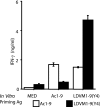
IFN-γ production in response to Ac1-9 or LDVM1-9. B10.PL mice were immunized with either Ac1-9 (open bars) or LDVM1-9(Y4) (closed bars) and draining lymph node cells were harvested and assayed for IFN-γ secretion when cultured with either medium, Ac1-9, or LDVM1-9(Y4). Culture with LDVM1-9(Y4) induced a strong IFN-γ response when mice were immunized with either Ac1-9 or LDVM1-9(Y4). In contrast, IFN-γ secretion was greatly reduced when animals were immunized with LDVM1-9(Y4) and draining lymph node cells were cultured with Ac1-9.
Fig. 5.
Fig. 5.
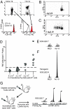
Demonstration of T cell competition using adoptive transfer strategies. (A) Splenocytes from Ac1-9-specific Vβ8.2Jβ2.4 and Vβ8.2Jβ2.7 transgenic mice were isolated and stained with PerCP-labeled anti-CD4 and either PE-labeled anti-TCRβ (H57-597) or PE-labeled, tetrameric MBP1-9[4Y]:I-Au complexes. Stained cells were analyzed by flow cytometry and data shown were gated on CD4+ T cells. (B) B10.PL animals received an adoptive transfer of 5 × 10⁶ CFSE-labeled CD4+ T cells obtained from the Vβ8.2Jβ2.4 Tg mouse. After the transfer, if the animals were not primed with Ac1-9 the T cells did not proliferate. (C) One day after transfer of 5 × 10⁶ Vβ8.2Jβ2.4 CFSE-labeled Tg T cells, animals were primed with Ac1-9, which resulted in proliferation of the transferred T cells. (D) Adoptive transfer of competing naïve Ac1-9-specific transgenic T cells effectively prevents the expansion of the public Vβ8.2Jβ2.7 driver clone. Mice received 0, 5 × 10³, 5 × 10⁴, and 5 × 10⁵ competing T cells isolated from the Vβ8.2Jβ2.4 Tg mouse. Twenty-four hours later, they were primed with 75 μg of Ac1-9 emulsified in CFA. The expansion of the endogenous Ac1-9-specific Vβ8.2Jβ2.7 clone was lost in a dose-dependent fashion. The strength of individual Vβ8.2Jβ2.7 expansions corresponding to a CDR3 length of 9 aa are shown numerically as a relative index of stimulation. (E) Actual Vβ8.2Jβ2.7 spectra seen after transfer of 5 × 10⁴ Vβ8.2Jβ2.4 Ac1-9-specific T cells. In the presence of competing T cells, the endogenous Vβ8.2Jβ2.7 clone failed to expand. (F) Vβ8.2Jβ2.4 spectra showing expansion of the adoptively transferred Vβ8.2Jβ2.4 T cells after immunization with Ac1-9. (G) Depletion of the exclusively LDVM1-9(Y4)-specific competing T cells allows the Ac1-9-specific Vβ8.2Jβ2.7 clonotype to expand following priming with LDVM1-9(Y4). Competing T cells were depleted via i.p. injection of anti-CD3 and anti-Vβ7 antibody. Following this depleting protocol mice were reconstituted with noncompeting T cells by adoptive transfer of unexpanded bulk Vβ8 T cells. Mice were finally immunized with 25 μg of LDVM1-9(Y4) and euthanized 10 days later for analysis. Following this regimen the naïve Vβ8.2Jβ2.7 Ac1-9-specific clonotype strongly responded to priming with LDVM1-9(Y4).

Similar articles

Cited by

References

    1. Bhardwaj V, Kumar V, Geysen HM, Sercarz EE. Degenerate recognition of a dissimilar antigenic peptide by myelin basic protein-reactive T cells. Implications for thymic education and autoimmunity. J Immunol. 1993;151:5000–5010. - PubMed
    1. Wucherpfennig KW, Strominger JL. Molecular mimicry in T cell-mediated autoimmunity: Viral peptides activate human T cell clones specific for myelin basic protein. Cell. 1995;80:695–705. - PMC - PubMed
    1. Hagerty DT, Allen PM. Intramolecular mimicry. Identification and analysis of two cross-reactive T cell epitopes within a single protein. J Immunol. 1995;155:2993–3001. - PubMed
    1. Hemmer B, et al. Identification of high potency microbial and self ligands for a human autoreactive class II-restricted T cell clone. J Exp Med. 1997;185:1651–1659. - PMC - PubMed
    1. Loftus C, Huseby E, Gopaul P, Beeson C, Goverman J. Highly cross-reactive T cell responses to myelin basic protein epitopes reveal a nonpredictable form of TCR degeneracy. J Immunol. 1999;162:6451–6457. - PubMed

Publication types

LinkOut - more resources