. 2010 Feb 16;107(7):2842-7.
doi: 10.1073/pnas.0914036107. Epub 2010 Jan 27.

Keap1 is a forked-stem dimer structure with two large spheres enclosing the intervening, double glycine repeat, and C-terminal domains

Affiliations

Keap1 is a forked-stem dimer structure with two large spheres enclosing the intervening, double glycine repeat, and C-terminal domains

Toshihiko Ogura et al. Proc Natl Acad Sci U S A. .

Abstract

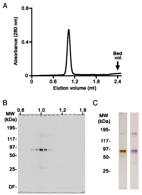
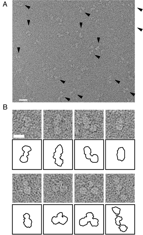
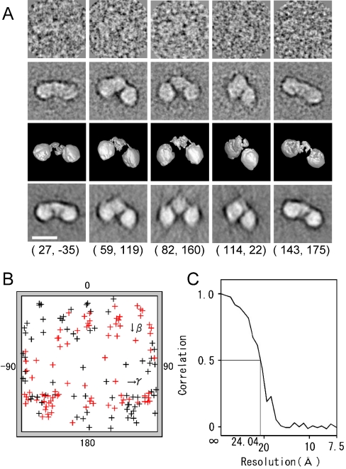
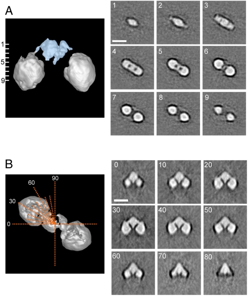
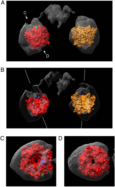
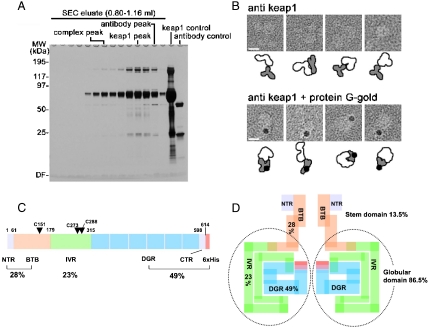
Keap1 is a substrate adaptor of a Cullin 3-based E3 ubiquitin ligase complex that recognizes Nrf2, and also acts as a cellular sensor for xenobiotics and oxidative stresses. Nrf2 is a transcriptional factor regulating the expression of cytoprotective enzyme genes in response to such stresses. Under unstressed conditions Keap1 binds Nrf2 and results in rapid degradation of Nrf2 through the proteasome pathway. In contrast, upon exposure to oxidative and electrophilic stress, reactive cysteine residues in intervening region (IVR) and Broad complex, Tramtrack, and Bric-à-Brac domains of Keap1 are modified by electrophiles. This modification prevents Nrf2 from rapid degradation and induces Nrf2 activity by repression of Keap1. Here we report the structure of mouse Keap1 homodimer by single particle electron microscopy. Three-dimensional reconstruction at 24-A resolution revealed two large spheres attached by short linker arms to the sides of a small forked-stem structure, resembling a cherry-bob. Each sphere has a tunnel corresponding to the central hole of the beta-propeller domain, as determined by x-ray crystallography. The IVR domain appears to surround the core of the beta-propeller domain. The unexpected proximity of IVR to the beta-propeller domain suggests that any distortions generated during modification of reactive cysteine residues in the IVR domain may send a derepression signal to the beta-propeller domain and thereby stabilize Nrf2. This study thus provides a structural basis for the two-site binding and hinge-latch model of stress sensing by the Nrf2-Keap1 system.


Conflict of interest statement

The authors declare no conflict of interest.

Figures

Fig. 1.
Fig. 1.
(A) Size exclusion chromatography. Recombinant Keap1 was purified using a Superdex S200 SEC column before EM analysis. Protein was routinely eluted at 1.03 mL as a single sharp peak; this fraction was used for EM image analysis. (B) SDS-PAGE and silver staining. The purity of fractions eluted from the SEC was monitored by SDS-PAGE and silver staining. (C) Purity of Keap1 was verified by silver staining (Left) and immunoblotting analysis using anti-Keap1 antibody (Right).
Fig. 2.
Fig. 2.
EM image of negatively stained Keap1. (A) Keap1 particles were observed as uniformly sized projections (arrowheads). Protein is shown in bright shades. Scale bar, 200 Å. (B) Examples of the Keap1 projections with schematic diagrams below each panel. An example of a minor population with extremely distant round spheres is also displayed at the rightmost bottom. Scale bar, 100 Å. For statistical analysis, 216 particles were automatically picked up by the auto-accumulation method and utilized as training data for the three-layer neural network (NN) auto-picking system. The trained NN selected 12,651 particles for analysis.
Fig. 3.
Fig. 3.
Three-dimensional reconstruction of Keap1. (A) Raw images of Keap1 with different Euler angles (Row 1), compared with the corresponding two-dimensional averages (Row 2), the surface views of the three-dimensional reconstruction (Row 3), and the reprojections of the three-dimensional reconstruction (Row 4) consistent through the reconstruction. The Euler angle (β, γ) is denoted below each column. Protein is displayed in bright shades. Scale bar, 100 Å. (B) Surface projection of Euler angle (β, γ) distribution of 162 adopted class averages. The distribution covers the whole angular range, with a small bias reflecting a somewhat preferred orientation of Keap1 molecules on the carbon surface. (C) Fourier shell correlation function indicates the resolution limit of 24 Å by the FSC > 0.5 criterion.
Fig. 4.
Fig. 4.
Sections through Keap1 molecule. (A) Horizontal slices perpendicular to the pseudo twofold symmetric axis show the characteristic cherry-shaped structure of Keap1 (Right). Positions of the cross sections, at 9.43 Å intervals throughout the molecule, are numbered from one to nine on the side surface view (Left). The stem structure, occupying 13.5% of the total volume (Blue). (B) Axial slices (Orange Dotted Lines; top surface view) every 10 ° from 0 ° to 90 ° (Left). Each sectional image is shown with corresponding angle (Right). Two globular domains are connected to the central stem-like density with slender linkers. The central stem-like density comprises two thin layers connected to one another but separated by a thin gap. The globular domains on either side are dense cylinders with round corners, pierced by an apparent low-density tunnel between the two surface orifices. Protein is displayed in bright shades. Scale bars, 100 Å.
Fig. 5.
Fig. 5.
Molecular fitting of the DC crystal structure to the Keap1 volume. (A) Atomic model of the Keap1-DC domain (PDB accession code 1X2J (15)), (Orange or Red), fitted into the EM density map. (B) Front half cutaway view of Keap1 molecule. One tunnel is inclined at 20 ° from the pseudosymmetry axis, while the other is almost parallel to the axis (White Dotted Lines). Tunnels closely coincide with those in the superimposed atomic model. (C) View from direction “C” as depicted in A. (D) View from direction “D” as depicted in A. The small exit of each tunnel closely coincides with that in the x-ray model. Extra density observed in the globular domain is composed of the IVR domain and part of the BTB domain, as described in Fig 6D. Distance between the two Nrf2 binding sites located on the bottom of the DC domains is approximately 80 Å. The N and C termini of the DC domain are denoted by Blue and Green, respectively.
Fig. 6.
Fig. 6.
BTB, IVR, and DC domains of Keap1.(A) Purification of the anti-DC antibody-bound Keap1 complex. SDS-PAGE analysis shows isolation of the antibody/Keap1 complex by SEC. The purified complex was eluted in early fractions at approximately 290-kDa region. (B) The large cherries of the molecule contain the DC domains. Gallery of negatively stained anti-DC/Keap1 complexes (Upper) and protein-G-gold/anti-DC antibody/Keap1 complexes (Bottom). Schematic diagrams of Keap1 (White), antibodies (Gray), and gold particles (Black) are illustrated below each panel. Scale bars,100 Å. (C) Protein architecture of Keap1 showing BTB, IVR, DGR, and CTR domains with corresponding primary sequence boundaries. Reactive cysteines (Cys151, Cys273, and Cys288) important for stress sensing are indicated with single letter codes and residue numbers. Percentages (%) of each functional domain are also shown. (D) Schematic distribution of the four functional domains in the Keap1 homodimer, as predicted by volume ratio. The stem structure occupies only 13.5% of the whole volume, while the globular regions comprise about 86.5%, suggesting that the entire IVR and part of the BTB domain are integrated into the globular cherries.

References

    1. Itoh K, et al. An Nrf2/small Maf heterodimer mediates the induction of phase II detoxifying enzyme genes through antioxidant response elements. Biochem Biophys Res Commun. 1997;236:313–322. - PubMed
    1. Itoh K, et al. Keap1 represses nuclear activation of antioxidant responsive elements by Nrf2 through binding to the amino-terminal Neh2 domain. Genes Dev. 1999;13:76–86. - PMC - PubMed
    1. Kobayashi M, Yamamoto M. Nrf2-Keap1 regulation of cellular defense mechanisms against electrophiles and reactive oxygen species. Adv Enzyme Regul. 2006;46:113–140. - PubMed
    1. Wakabayashi N, et al. Keap1-null mutation leads to postnatal lethality due to constitutive Nrf2 activation. Nat Genet. 2003;35:238–245. - PubMed
    1. Okawa H, et al. Hepatocyte-specific deletion of the keap1 gene activates Nrf2 and confers potent resistance against acute drug toxicity. Biochem Biophys Res Commun. 2006;339:79–88. - PubMed

Publication types

MeSH terms

Substances