. 2010 Feb 16;107(7):3146-51.
doi: 10.1073/pnas.0910717107. Epub 2010 Jan 26.

Hsp90 inhibitors block outgrowth of EBV-infected malignant cells in vitro and in vivo through an EBNA1-dependent mechanism

Affiliations

Hsp90 inhibitors block outgrowth of EBV-infected malignant cells in vitro and in vivo through an EBNA1-dependent mechanism

Xiaoping Sun et al. Proc Natl Acad Sci U S A. .

Abstract

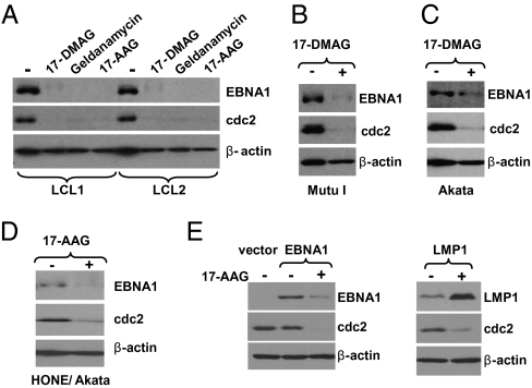
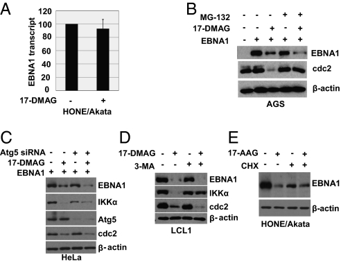
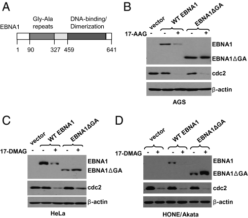
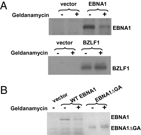
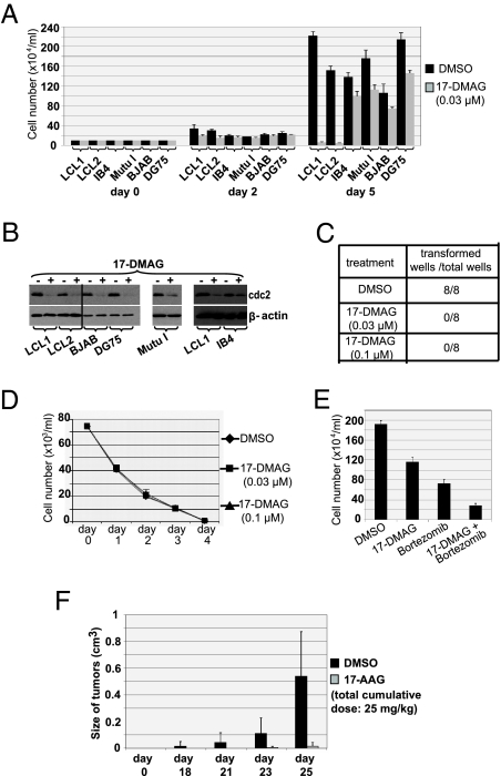
EBV causes infectious mononucleosis and is associated with certain malignancies. EBV nuclear antigen 1 (EBNA1) mediates EBV genome replication, partition, and transcription, and is essential for persistence of the viral genome in host cells. Here we demonstrate that Hsp90 inhibitors decrease EBNA1 expression and translation, and that this effect requires the Gly-Ala repeat domain of EBNA1. Hsp90 inhibitors induce the death of established, EBV-transformed lymphoblastoid cell lines at doses nontoxic to normal cells, and this effect is substantially reversed when lymphoblastoid cell lines are stably infected with a retrovirus expressing a functional EBNA1 mutant lacking the Gly-Ala repeats. Hsp90 inhibitors prevent EBV transformation of primary B cells, and strongly inhibit the growth of EBV-induced lymphoproliferative disease in SCID mice. These results suggest that Hsp90 inhibitors may be particularly effective for treating EBV-induced diseases requiring the continued presence of the viral genome.


Conflict of interest statement

The authors declare no conflict of interest.

Figures

Fig. 1.
Fig. 1.
Hsp90 inhibitors decrease expression of EBNA1. (A) LCL cells (line 1 and 2) were treated with no drug, 17-DMAG (0.17 μM), geldanamycin (0.5 μM), or 17-AAG (0.5 μM) for 96 h. (B and C) Mutu I and Akata Burkitt lymphoma cells were treated with no drug or 17-DMAG (0.17 μM) for 96 h. (D) The EBV-infected NPC cell line HONE/Akata was treated with no drug or 17-AAG (0.5 μM) for 48 h. (E) AGS gastric cells (EBV-negative) were transfected with empty vector (pSG5), pSG5-EBNA1, or pSG5-LMP1 as indicated, followed by a 48-h treatment with 17-AAG (0.25 μM) beginning at 4 h after transfection. Whole-cell extracts were prepared and immunoblot analysis was performed to analyze the expression of EBNA1, cdc2 (a known cellular substrate of Hsp90), cellular β-actin, or LMP1 as indicated.
Fig. 2.
Fig. 2.
Hsp90 inhibitors reduce EBNA1 independent of effects on EBNA1 transcripts or EBNA1 stability. (A) EBV-positive HONE/Akata cells were treated with 17-DMAG (0.17 μM) for 24 h. Expression of the EBNA1 transcript was examined by quantitative RT-PCR. The level of EBNA1 transcript in untreated cells is set as 100. (B) EBV-negative AGS cells were transfected with empty vector (pSG5) or pSG5-EBNA1, followed by a 48-h treatment with no drug or 17-DMAG (0.17 μM) beginning at 4 h after transfection in the presence or absence of the proteasome inhibitor MG-132 (50 μM). (C) HeLa cells were transfected with pSG5-EBNA1 in the presence of Atg5 siRNA or equivalent amounts of a control siRNA, then treated with no drug or 17-DMAG (0.17 μM). (D) LCL1 cells were treated with no drug or 17-DMAG (0.17 μM) in the presence or absence of the autophagy inhibitor 3-MA (10 mM). (E) EBV-positive HONE/Akata cells were treated with no drug or 17-AAG (0.5 μM) for 48 h in the presence or absence of CHX (50 μg/mL) added into medium 12 h before cell harvesting. Whole-cell extracts were prepared and immunoblot analysis was performed to analyze the expression of viral and cellular proteins as indicated (BE).
Fig. 3.
Fig. 3.
The Gly-Ala repeats are required for inhibition of EBNA1 expression by Hsp90 inhibitors. (A) Schematic diagram of the EBNA1 protein showing some of its functional elements and amino acid numbers. AGS cells (B), HeLa cells (C), and EBV-positive HONE cells (D) were transfected with empty vector (pSG5), pSG5-EBNA1, or pSG5-EBNA1ΔGA (a mutant protein missing the Gly-Ala repeats), followed by a 48-h treatment with 17-AAG (0.5 μM) or 17-DMAG (0.17 μM) beginning at 4 h after transfection. Immunoblot analysis was performed to analyze the expression of EBNA1, cdc2, and β-actin.
Fig. 4.
Fig. 4.
Geldanamycin inhibits EBNA1 translation in reticulocyte lysate. (A) pSG5 expression constructs encoding EBNA1 and BZLF1 were transcribed and translated in vitro with T7 RNA polymerase using a coupled transcription–translation reticulocyte lysate system supplemented with [35S]methionine/cysteine in the presence or absence of geldanamycin (0.5 μM). EBNA1 translation was reduced by 74% in the presence of geldanamycin. (B) pcDNA3.1 expression constructs encoding EBNA1 and EBNA1ΔGA were also transcribed and translated in vitro using the same method in the presence or absence of geldanamycin (0.5 μM). Ten fold less EBNA1ΔGA versus EBNA1 protein is loaded on the gel.
Fig. 5.
Fig. 5.
Hsp90 inhibitors reduce viability of EBV-transformed LCLs and prevent EBV transformation of primary B cells. (A) Two different independently derived LCLs (late passage line LCL1 and early passage line LCL2), IB4 (an LCL in which the EBV genome is integrated), two EBV-negative B-cell lymphoma lines (BJAB and DG75), and an EBV+ Burkitt lymphoma line (Mutu I), were treated with either vehicle control (DMSO) or 17-DMAG (0.03 μM). Cell counts in each condition were determined by trypan blue exclusion at the time points indicated. (B) Whole-cell extracts were prepared from cells treated with or without 17-DMAG (0.17 μM) and immunoblot analysis was performed to analyze the expression of cellular proteins as indicated. (C) Primary B cells were infected with EBV and treated with either vehicle control (DMSO) or 17-DMAG (0.03 μM or 0.1 μM) beginning 1 h after infection. Media (and drug) were replaced once per week. Numbers of transformed wells and the total numbers of wells treated with either no drug (DMSO) or 17-DMAG (0.03 μM or 0.1 μM) at 3 weeks after infection are given. (D) Primary B cells were treated with either no drug or 17-DMAG (0.03 μM or 0.1 μM). Cell counts in each condition were determined by trypan blue exclusion at the time points as indicated. (E) LCL1 cells were treated with no drug, 17-DMAG (0.005 μM), bortezomib (0.01 μM), or both, and cell counts determined. (F) EBV-positive lymphoblastoid cells (5 × 10⁶ LCL1 cells) were implanted s.c. into the flanks of SCID mice. Tumors (six mice in each group) were treated with either no drug or three low doses (total cumulative dose, 25 mg/kg) of 17-AAG (given i.p. on d 7, 9, and 11 after injection of LCL1 cells). Columns show mean tumor volumes at different time points; bars show SE.
Fig. 6.
Fig. 6.
Expression of an EBNA1 mutant missing the Gly-Ala repeat domain decreases the toxic effect of Hsp90 inhibitors in LCLs. (A) LCL1 cells were infected with a retrovirus vector expressing EBNA1ΔGA or the control retrovirus vector. Immunoblot analysis was performed to analyze the expression of EBNA1, EBNA1ΔGA, cdc2, and β-actin in the presence and absence of 17-DMAG. (B) LCL1-vector and LCL1-EBNA1ΔGA cells were treated with either no drug or 17-DMAG (0.03 μM or 0.1 μM). Cell counts in each condition were determined by trypan blue exclusion at the time points indicated. (C) LCL1-vector and LCL1-EBNA1ΔGA cells were treated with either no drug or MTX (0.1 μg/mL or 0.5 μg/mL). Cell counts in each condition were determined by trypan blue exclusion at the time points indicated. (D) LCL1-vector and LCL1-EBNA1ΔGA cells were treated with either no drug or 17-DMAG (0.03 μM) for 48 h. The cell cycle distribution is given.

Similar articles

Cited by

References

    1. Kieff E, Richinson AB. 2007. Epstein-Barr virus and its replication. Fields’ virology. (Lippincott Williams & Wilkins, Philadelphia) 5th ed. eds Knipe D, etal., pp 2603–2654.
    1. Niller HH, Wolf H, Minarovits J. Regulation and dysregulation of Epstein-Barr virus latency: implications for the development of autoimmune diseases. Autoimmunity. 2008;41:298–328. - PubMed
    1. Lindner SE, Sugden B. The plasmid replicon of Epstein-Barr virus: mechanistic insights into efficient, licensed, extrachromosomal replication in human cells. Plasmid. 2007;58:1–12. - PMC - PubMed
    1. Kapoor P, Lavoie BD, Frappier L. EBP2 plays a key role in Epstein-Barr virus mitotic segregation and is regulated by aurora family kinases. Mol Cell Biol. 2005;25:4934–4945. - PMC - PubMed
    1. Wu H, Ceccarelli DF, Frappier L. The DNA segregation mechanism of Epstein-Barr virus nuclear antigen 1. EMBO Rep. 2000;1:140–144. - PMC - PubMed

Publication types

MeSH terms

LinkOut - more resources