. 2010 Feb 9;107(6):2443-8.
doi: 10.1073/pnas.0911235107. Epub 2010 Jan 21.

The protein tyrosine phosphatases PTPRZ and PTPRG bind to distinct members of the contactin family of neural recognition molecules

Affiliations

The protein tyrosine phosphatases PTPRZ and PTPRG bind to distinct members of the contactin family of neural recognition molecules

Samuel Bouyain et al. Proc Natl Acad Sci U S A. .

Abstract

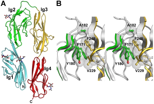
The receptor protein tyrosine phosphatases gamma (PTPRG) and zeta (PTPRZ) are expressed primarily in the nervous system and mediate cell adhesion and signaling events during development. We report here the crystal structures of the carbonic anhydrase-like domains of PTPRZ and PTPRG and show that these domains interact directly with the second and third immunoglobulin repeats of the members of the contactin (CNTN) family of neural recognition molecules. Interestingly, these receptors exhibit distinct specificities: PTPRZ binds only to CNTN1, whereas PTPRG interacts with CNTN3, 4, 5, and 6. Furthermore, we present crystal structures of the four N-terminal immunoglobulin repeats of mouse CNTN4 both alone and in complex with the carbonic anhydrase-like domain of mouse PTPRG. In these structures, the N-terminal region of CNTN4 adopts a horseshoe-like conformation found also in CNTN2 and most likely in all CNTNs. This restrained conformation of the second and third immunoglobulin domains creates a binding site that is conserved among CNTN3, 4, 5, and 6. This site contacts a discrete region of PTPRG composed primarily of an extended beta-hairpin loop found in both PTPRG and PTPRZ. Overall, these findings implicate PTPRG, PTPRZ and CNTNs as a group of receptors and ligands involved in the manifold recognition events that underlie the construction of neural networks.


Conflict of interest statement

The authors declare no conflict of interest.

Figures

Fig. 1.
Fig. 1.
Structures of CA domains from type V RPTPs. (A) Ribbon diagram of human PTPRZCA. The letters N and C indicate the N- and C-termini, respectively. Disulfide bonds are shown as orange ball-and-stick models. (B) Human PTPRGCA. The dotted line indicates a disordered β-hairpin loop. (C) Mouse PTPRGCA. (D) Overlay of the structures of human PTPRZCA (blue), human PTPRGCA (brown), and mouse PTPRGCA (magenta) with the structure of mouse CA-XIV (gray). A gray sphere indicates the position of the zinc ion in CA-XIV.
Fig. 2.
Fig. 2.
Affinity isolation of GPI-anchored CNTNs. CNTNs were fused to hGH and expressed transiently in HEK293 cells. Cell lysates were incubated with PTPRZCA-resin (A) or with PTPRGCA-resin (B). Bound CNTN fusion proteins were visualized by immunoblotting against hGH.
Fig. 3.
Fig. 3.
Interactions between PTPRZCA, PTPRGCA and fragments of mouse CNTNs. (AC) Fragments of mouse CNTNs were fused to hGH and expressed transiently in HEK293 cells. Conditioned media were incubated with PTPRZCA-resin (A) or with PTPRGCA-resin (B and C). Bound CNTN fusion proteins were visualized by immunoblotting against hGH. (D) PTPRZCA forms a 1∶1 complex with CNTN1Ig2-3. Comparison of size exclusion chromatograms of CNTN1Ig2-3, PTPRZCA, and CNTN1Ig2-3 mixed with PTPRZCA. Arrows above the chromatogram indicate the elution volumes of molecular weight standards. The insert shows fractions from the complex elution profile resolved by SDS-PAGE. The proteins were visualized by silver staining. (E) PTPRGCA forms a 1∶1 complex with CNTN4Ig1-4. Same as (D), but the experiments were conducted with CNTN4Ig1-4 and PTPRGCA.
Fig. 4.
Fig. 4.
Structure of mouse CNTN4Ig1-4. (A) Ribbon diagram of mouse CNTN4Ig1-4. The letters N and C indicate the N- and C-termini, respectively. Disulfide bonds are shown as orange ball-and-stick models. Asparagine-linked N-acetylglucosamine residues are depicted as gray ball-and-stick models along with the asparagine side chain. Ig domains 1, 2, 3, and 4 are colored cyan, green, gold, and red, respectively. (B) Stereo view of the interface between Ig domains 2 and 3 in CNTN4Ig1-4. Residues at the interface between the two domains are shown as ball-and-sticks with transparent spheres (gray) and colored green (Ig2) or gold (Ig3).
Fig. 5.
Fig. 5.
Structure of the PTPRGCA·CNTN4Ig1-4 complex. The view on the left is obtained from the one shown in Fig. 4 A by a counterclockwise rotation of approximately 60° along a vertical axis. In the right view, only residues 225–229 and 288–301 (β-hairpin) of PTPRGCA are shown for the sake of clarity. The letters N and C indicate the N- and C-termini, respectively. Disulfide bonds are shown as orange ball-and-stick models. Asparagine-linked N-acetylglucosamine residues are depicted as gray ball-and-stick models along with the asparagine side chain. Ig domains 1, 2, 3, and 4 are colored cyan, green, gold, and red, respectively. PTPRGCA is colored magenta. Dotted lines indicate disordered regions. The β-hairpin region is well ordered with average B factors of 32.1  2 versus 33.8  2 for the entire chain of PTPRGCA.
Fig. 6.
Fig. 6.
Stereo view of the PTPRGCA·CNTN4Ig1-4 interface. This view is in the same orientation as the right view in Fig. 5. Residues are shown as ball-and-sticks with transparent gray spheres for those involved in van der Waals contacts. Dashed lines indicate potential hydrogen bonds and salt bridges. Residues from CNTN4Ig2, CNTN4Ig3, and PTPRGCA are colored green, gold, and magenta, respectively.

Similar articles

Cited by

References

    1. Johnson KG, Van Vactor D. Receptor protein tyrosine phosphatases in nervous system development. Physiol Rev. 2003;83:1–24. - PubMed
    1. Andersen JN, et al. Structural and evolutionary relationships among protein tyrosine phosphatase domains. Mol Cell Biol. 2001;21:7117–7136. - PMC - PubMed
    1. Harroch S, et al. No obvious abnormality in mice deficient in receptor protein tyrosine phosphatase beta. Mol Cell Biol. 2000;20:7706–7715. - PMC - PubMed
    1. Lamprianou S, et al. Receptor protein tyrosine phosphatase gamma is a marker for pyramidal cells and sensory neurons in the nervous system and is not necessary for normal development. Mol Cell Biol. 2006;26:5106–5119. - PMC - PubMed
    1. Tripp BC, Smith K, Ferry JG. Carbonic anhydrase: new insights for an ancient enzyme. J Biol Chem. 2001;276:48615–48618. - PubMed

MeSH terms