

Analysis of the cellular mechanism underlying inhibition of EAE after treatment with anti-NKG2A F(ab')2
- PMID: 20133787
- PMCID: PMC2823885
- DOI: 10.1073/pnas.0914732107
Analysis of the cellular mechanism underlying inhibition of EAE after treatment with anti-NKG2A F(ab')2
Abstract
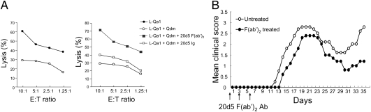
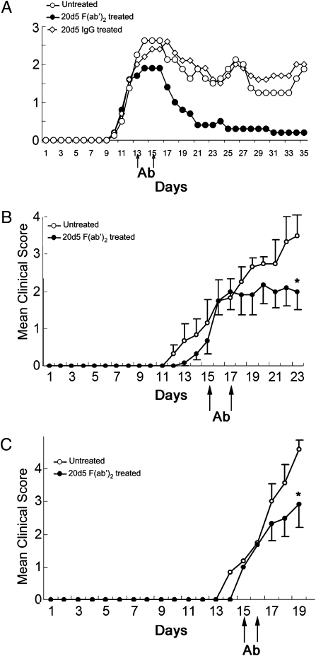
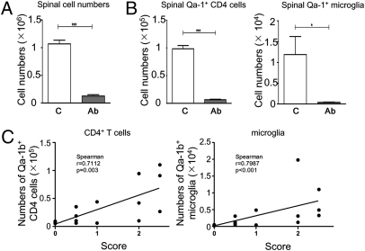
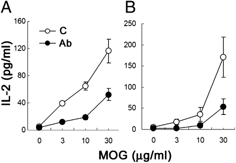
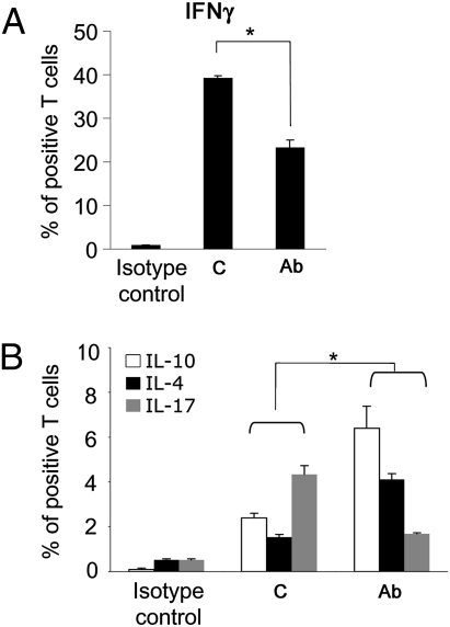
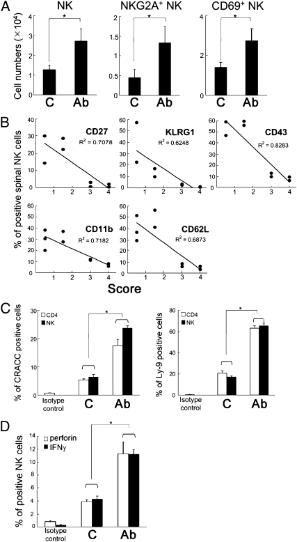
Autoimmune encephalomyelitis may be ameliorated experimentally by enhancing NK cell-mediated elimination of activated autoreactive T cells through a mutation that interrupts the interaction between Qa-1(b) and CD94/NKG2A. Here we evaluate the ability of an anti-NKG2A F(ab')(2) Ab to enhance elimination of autoreactive T cells and reduce experimental autoimmune encephalomyelitis (EAE). Anti-NKG2A F(ab')(2) treatment diminishes progression of both myelin oligodendrocyte glycoprotein (MOG)-induced EAE in intact C57BL/6 mice and after adoptive transfer of disease-causing T cells. Analyses of the underlying mechanism revealed that administration of anti-NKG2A F(ab')(2) Ab reduces CD4(+) T recall responses to MOG and skews the proportion of IL-17- and IFNgamma-producing CD4(+) T cells toward the protective IL-4- and IL-10-secreting CD4(+) T cell subpopulations. CD94/NKG2A-dependent inhibition of inflammatory damage to spinal cord is associated with decreased infiltration of T cells and reduced microglia activation in the central nervous system. Because anti-NKG2A F(ab')(2) treatment had no detectable effect on the numbers or activity of T and B lymphocytes and NK cells in peripheral lymphoid tissues, this anti-NKG2A-based approach may represent a safe and effective therapy for this CNS disorder.
Conflict of interest statement
The authors declare no conflict of interest.
Figures
Similar articles
-
Infection with Mycobacterium bovis BCG diverts traffic of myelin oligodendroglial glycoprotein autoantigen-specific T cells away from the central nervous system and ameliorates experimental autoimmune encephalomyelitis.Clin Diagn Lab Immunol. 2003 Jul;10(4):564-72. doi: 10.1128/cdli.10.4.564-572.2003. Clin Diagn Lab Immunol. 2003. PMID: 12853387 Free PMC article.
-
Regulation of experimental autoimmune encephalomyelitis in the C57BL/6J mouse by NK1.1+, DX5+, alpha beta+ T cells.J Immunol. 2001 Mar 15;166(6):4209-15. doi: 10.4049/jimmunol.166.6.4209. J Immunol. 2001. PMID: 11238673
-
Regulation of activated CD4+ T cells by NK cells via the Qa-1-NKG2A inhibitory pathway.Immunity. 2007 May;26(5):593-604. doi: 10.1016/j.immuni.2007.03.017. Immunity. 2007. PMID: 17509909 Free PMC article.
-
RTL551 treatment of EAE reduces CD226 and T-bet+ CD4 T cells in periphery and prevents infiltration of T-bet+ IL-17, IFN-γ producing T cells into CNS.PLoS One. 2011;6(7):e21868. doi: 10.1371/journal.pone.0021868. Epub 2011 Jul 5. PLoS One. 2011. PMID: 21750737 Free PMC article.
-
Monalizumab: inhibiting the novel immune checkpoint NKG2A.J Immunother Cancer. 2019 Oct 17;7(1):263. doi: 10.1186/s40425-019-0761-3. J Immunother Cancer. 2019. PMID: 31623687 Free PMC article. Review.
Cited by
-
Implications of NKG2A in immunity and immune-mediated diseases.Front Immunol. 2022 Aug 10;13:960852. doi: 10.3389/fimmu.2022.960852. eCollection 2022. Front Immunol. 2022. PMID: 36032104 Free PMC article. Review.
-
NK Cells in Autoimmune Diseases: Protective or Pathogenic?Front Immunol. 2021 Mar 12;12:624687. doi: 10.3389/fimmu.2021.624687. eCollection 2021. Front Immunol. 2021. PMID: 33777006 Free PMC article. Review.
-
Natural killer cells and their receptors in multiple sclerosis.Brain. 2013 Sep;136(Pt 9):2657-76. doi: 10.1093/brain/aws159. Epub 2012 Jun 25. Brain. 2013. PMID: 22734127 Free PMC article. Review.
-
Interleukin-2/interleukin-2 antibody therapy induces target organ natural killer cells that inhibit central nervous system inflammation.Ann Neurol. 2011 Apr;69(4):721-34. doi: 10.1002/ana.22339. Epub 2011 Mar 18. Ann Neurol. 2011. PMID: 21425186 Free PMC article.
-
Regulatory Functions of Natural Killer Cells in Multiple Sclerosis.Front Immunol. 2016 Dec 19;7:606. doi: 10.3389/fimmu.2016.00606. eCollection 2016. Front Immunol. 2016. PMID: 28066417 Free PMC article. Review.
References
-
- Lublin FD. Adoptive transfer of murine relapsing experimental allergic encephalomyelitis. Ann Neurol. 1985;17:188–190. - PubMed
-
- Noseworthy JH, Lucchinetti C, Rodriguez M, Weinshenker BG. Multiple sclerosis. N Engl J Med. 2000;343:938–952. - PubMed
-
- Yednock TA, et al. Prevention of experimental autoimmune encephalomyelitis by antibodies against α 4 β 1 integrin. Nature. 1992;356:63–66. - PubMed
-
- Han MH, et al. Proteomic analysis of active multiple sclerosis lesions reveals therapeutic targets. Nature. 2008;451:1076–1081. - PubMed
Publication types
MeSH terms
Substances
Grants and funding
LinkOut - more resources
Full Text Sources
Other Literature Sources
Research Materials
