. 2010 Feb 9;107(6):2586-91.
doi: 10.1073/pnas.0913054107. Epub 2010 Jan 25.

Evidence for a functional role of IgE anticitrullinated protein antibodies in rheumatoid arthritis

Affiliations

Evidence for a functional role of IgE anticitrullinated protein antibodies in rheumatoid arthritis

A J M Schuerwegh et al. Proc Natl Acad Sci U S A. .

Retraction in

Abstract

Rheumatoid arthritis (RA) is a systemic autoimmune disease involving inflammation of the joints. Among the autoantibodies described in RA, anticitrullinated protein antibodies (ACPAs) are highly specific and predictive for RA. In addition, ACPAs have been implicated in the pathogenesis of RA. However, a direct functional response of immune cells from ACPA(+) RA patients toward citrullinated proteins has not been demonstrated. In this study, we show that exposure to citrullinated antigens leads to activation of basophils from ACPA(+) RA patients within 20 minutes. This was not observed after exposure of basophils to noncitrullinated control antigens or after stimulation of basophils from ACPA(-) RA patients and healthy controls. Basophil activation was correlated with the binding of citrullinated proteins to basophils. Furthermore, serum from ACPA(+) RA patients in contrast to that from ACPA(-) RA patients could specifically sensitize human FcepsilonRI expressing rat basophil cells (RBL), enabling activation by citrullinated proteins. Mast cell degranulation products such as histamine levels were enhanced in synovial fluid of ACPA(+) RA patients as compared with ACPA(-) RA and osteoarthritis patients. In addition, histamine levels in synovial fluid from ACPA(+) RA patients correlated with IgE levels, suggesting degranulation of mast cells by cross-linking IgE. Immunohistochemistry on synovial biopsies demonstrated an increased number of degranulated CD117(+) mast cells in ACPA(+) RA patients; IgE and FcepsilonRI expression in synovial mast cells from ACPA(+) RA patients was increased. In conclusion, our results show an immunological response of immune cells from ACPA(+) RA patients in a citrulline-specific manner. Moreover, these data indicate a role for IgE-ACPAs and FcepsilonRI-positive cells in the pathogenesis of RA.


Conflict of interest statement

The authors declare no conflict of interest.

Figures
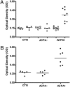
Fig. 1.
Fig. 1.
IgE-ACPAs. (A) Anti-IgE antibodies against noncitrullinated fibrinogen (NC-FB) (○) and citrullinated fibrinogen (C-FB) (●) in IgG-depleted serum samples from control (CTR; n = 5), ACPA RA (n = 4), and ACPA+ RA (n = 8) patients. (B) Anti-IgE-ACPAs in IgG-depleted serum samples from CTR (n = 5), ACPA RA (n = 4), and ACPA+ RA (n = 6) patients. P = 0.003 in ACPA+ RA vs. CTR. P = 0.007 in ACPA RA vs. ACPA+ RA.
Fig. 2.
Fig. 2.
Basophil activation. Whole heparinized blood was stimulated with HBSS buffer, 1 μg/mL anti-immunoglobulin E (anti-IgE), 0.2 U/mL PAD, 10 μg/mL noncitrullinated fibrinogen (NC-FB), and 10 μg/mL citrullinated fibrinogen (C-FB) in control (CTR; n = 26), ACPA RA (n = 15), and ACPA+ RA (n = 29) patients. Bars represent median values. P < 0.0001 for HBSS vs. aIgE in CTR, ACPA RA, and ACPA+ RA. P < 0.0001 for C-FB vs. NC-FB in ACPA+ RA. P < 0.0001 for C-FB in ACPA+ RA vs. ACPA RA. P < 0.0001 for C-FB in ACPA+ RA vs. CTR.
Fig. 3.
Fig. 3.
Basophil activation with citrullinated (C)-MBP, heat-aggregated IgG (HAIgG), and IgG. Whole heparinized blood was stimulated with HBSS buffer, 1 μg/mL anti-IgE, 10 μg/mL noncitrullinated (NC)-MBP, 10 C-MBP, 10 μg/mL NC-fibrinogen (FB), 10 μg/mL C-FB, 10 μg/mL monomeric IgG, and 10 or 100 μg/mL HAIgG in healthy control (CTR; n = 3), ACPA RA (n = 2), and ACPA+ RA (n = 6) patients. P = 0.02 in ACPA+ RA vs. CTR and ACPA RA.
Fig. 4.
Fig. 4.
Elution of IgG. Basophil activation before and after elution of Ig with acid glycine buffer in ACPA+ RA patients (n = 5). Basophils were stimulated with HBSS buffer, 1 μg/mL anti-IgE, 10 μg/mL noncitrullinated fibrinogen (NC-FB), 10 μg/mL citrullinated fibrinogen (C-FB), and 1 μg/mL fMLP before and after elution of Ig with acid glycine buffer in ACPA+ RA patients (n = 5). P = 0.04 for C-FB PRE elution vs. C-FB POST elution.
Fig. 5.
Fig. 5.
Passive sensitization of FcεRI-transfected RBLs. After sensitization with sera from control (CTR; n = 10), ACPA RA (n = 39), and ACPA+ RA (n = 50) patients for 18 hours, RBLs were stimulated with Thyrod’s buffer, 1 μg/mL anti-IgE, 10 μg/mL noncitrullinated fibrinogen (NC-FB), or 10 μg/mL citrullinated fibrinogen (C-FB). Results are expressed as the percentage of total β-hexoaminidase release (treated with Triton-X) after correction for spontaneous release in Thyrod’s buffer. P < 0.0001 for C-FB vs. NC-FB in ACPA+ RA. P < 0.0001 for C-FB in ACPA+ RA vs ACPA RA. P = 0.001 for C-FB in ACPA+ RA vs CTR.
Fig. 6.
Fig. 6.
Surface IgE expression on CD117-allophycocyanin (APC+) synovial mast cells. Synovial mast cells of ACPA+ RA (n = 10), ACPA RA (n = 5), and OA (n = 18) patients were stained with Alexafluor-conjugated anti-IgE. Results are expressed as difference of mean fluorescence intensity (MFI) between isotype control MFI and anti-IgE MFI. P = 0.008 in ACPA+ RA vs. ACPA RA. P < 0.0001 in ACPA+ RA vs. OA.
Fig. 7.
Fig. 7.
Histamine levels in RA synovial fluid. Histamine levels (ng/mL) in synovial fluid of OA (n = 14), ACPA RA (n = 36), and ACPA+ RA (n = 39) patients. Bars represent median values. P = 0.004 in ACPA+ RA vs. OA. P = 0.008 in ACPA RA+ vs. ACPA RA.
Fig. 8.
Fig. 8.
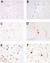
Microscopic features of the synovial tissue in ACPA RA and ACPA+ RA. (A) CD117 staining of mast cells. (Magnification: ×200.) (B) Staining of surface CD117 on mast cells. (Magnification: ×400.) (C) CAE staining of mast cell. (Magnification: ×400.) (D) CAE staining of granules in mast cells. (Magnification: ×1,000.) (E) Double CAE/ CD117 staining (whole granules not visible after antigen retrieval). (Magnification: ×400.) (F) Nondegranulated mast cell positive for CAE and CD117 (Left) and degranulated mast cell positive for CD117 and negative for CAE (Right). (Magnification: ×1,000.)

Similar articles

Cited by

References

    1. Schellekens GA, et al. The diagnostic properties of rheumatoid arthritis antibodies recognizing a cyclic citrullinated peptide. Arthritis Rheum. 2000;43:155–163. - PubMed
    1. van Gaalen FA, et al. Autoantibodies to cyclic citrullinated peptides predict progression to rheumatoid arthritis in patients with undifferentiated arthritis: A prospective cohort study. Arthritis Rheum. 2004;50:709–715. - PubMed
    1. Meyer O, et al. Anticitrullinated protein/peptide antibody assays in early rheumatoid arthritis for predicting five year radiographic damage. Ann Rheum Dis. 2003;62:120–126. - PMC - PubMed
    1. van der Helm-van Mil AH, Verpoort KN, Breedveld FC, Toes RE, Huizinga TW. Antibodies to citrullinated proteins and differences in clinical progression of rheumatoid arthritis. Arthritis Res Ther. 2005;7:R949–R958. - PMC - PubMed
    1. Kuhn KA, et al. Antibodies against citrullinated proteins enhance tissue injury in experimental autoimmune arthritis. J Clin Invest. 2006;116:961–973. - PMC - PubMed

Publication types

MeSH terms