. 2010 Feb 9;107(6):2568-73.
doi: 10.1073/pnas.0915000107. Epub 2010 Jan 25.

Two physically, functionally, and developmentally distinct peritoneal macrophage subsets

Affiliations

Two physically, functionally, and developmentally distinct peritoneal macrophage subsets

Eliver Eid Bou Ghosn et al. Proc Natl Acad Sci U S A. .

Abstract

The peritoneal cavity (PerC) is a unique compartment within which a variety of immune cells reside, and from which macrophages (MØ) are commonly drawn for functional studies. Here we define two MØ subsets that coexist in PerC in adult mice. One, provisionally called the large peritoneal MØ (LPM), contains approximately 90% of the PerC MØ in unstimulated animals but disappears rapidly from PerC following lipopolysaccharide (LPS) or thioglycolate stimulation. These cells express high levels of the canonical MØ surface markers, CD11b and F4/80. The second subset, referred to as small peritoneal MØ (SPM), expresses substantially lower levels of CD11b and F4/80 but expresses high levels of MHC-II, which is not expressed on LPM. SPM, which predominates in PerC after LPS or thioglycolate stimulation, does not derive from LPM. Instead, it derives from blood monocytes that rapidly enter the PerC after stimulation and differentiate to mature SPM within 2 to 4 d. Both subsets show clear phagocytic activity and both produce nitric oxide (NO) in response to LPS stimulation in vivo. However, their responses to LPS show key differences: in vitro, LPS stimulates LPM, but not SPM, to produce NO; in vivo, LPS stimulates both subsets to produce NO, albeit with different response patterns. These findings extend current models of MØ heterogeneity and shed new light on PerC MØ diversity, development, and function. Thus, they introduce a new context for interpreting (and reinterpreting) data from ex vivo studies with PerC MØ.


Conflict of interest statement

The authors declare no conflict of interest.

Figures
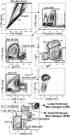
Fig. 1.
Fig. 1.
Naive mouse PerC contains a variety of immune cell subsets. Gating strategy used to identify immune cells present in PerC based on surface molecules. Total PerC cells from unstimulated BALB/c mice were processed and analyzed as described in Materials and Methods. Data shown are representative of >10 experiments.
Fig. 2.
Fig. 2.
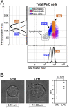
The two distinct PerC MØ subsets are differentiated by size. (A) PerC MØ (and other cell types) were analyzed for their scatter profile (forward and side: size and granularity, respectively) by flow cytometry. (B) SPMs and LPMs were FACS-sorted and their diameter were measured. Representative differential interference contrast images are shown.
Fig. 3.
Fig. 3.
SPMs and LPMs are absent in mouse blood. PerC and blood cells from unstimulated BALB/c mice were stained and analyzed for specific surface markers. *SPM and LPM were identified as in Fig. 1; blood monocytes were identified as Ly-6Chi, CD11b+ cells. SPMs and LPMs are also absent in other naive lymphoid organs, such as spleen and lymph nodes (Fig. S2).
Fig. 4.
Fig. 4.
Both SPMs and LPMs are able to phagocyte bacteria in vivo. E. coli (5 × 10⁶) expressing GFP were injected i.p. into BALB/c mice and, after 2 h, PerC cells were harvested and stained as described in Materials and Methods. Flow cytometry analysis reveals that most SPMs and LPMs were positive for GFP, indicating that they had internalized bacteria. Confocal images: GFP+ SPMs and LPMs were FACS-sorted and analyzed to confirm whether the bacteria had been internalized: green, internalized E. coli; blue, actin (phalloidin); red, anti-LPS (red staining indicates that bacteria have remained adherent to the outer cell membrane, i.e., not phagocytized).
Fig. 5.
Fig. 5.
SPMs and LPMs respond differently to TLR agonists. (A) PerC cells (106) were incubated with or without 1 μg/mL of LPS for 20 h at physiologic oxygen levels (5% O2). The specificity of the NO measurement was confirmed by adding 1.5 mM L-NAME reagent into the culture, which specifically inhibits NO formation. (B) BALB/c mice were injected i.p. with 5 μg of LPS (or PBS solution control) for 20 h. SPMs and LPMs were identified as in Fig. 1 and Fig. S1. NO production was measured by flow cytometry using DAF-FM diacetate. N/S, not significant. For additional control data, see Fig. S5.
Fig. 6.
Fig. 6.
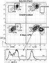
LPS induces a dramatic shift in the SPM:LPM ratio. BALB/c were injected i.p. with 10 μg of LPS. After 2 d, PerC cells were harvested, stained, and analyzed for the presence of myeloid cells. SPMs and LPMs were identified as described in Fig. 1. Neutrophils and eosinophils were best gated out from this analysis by using the gating strategy shown in Fig. S1.
Fig. 7.
Fig. 7.
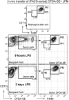
SPMs are developmentally independent from LPMs. FACS-sorted and CFDA-SE-labeled LPMs (2 × 10⁵) were injected i.p. into BALB/c recipients. Recipient mice were injected i.p. with LPS and, after 9 h or 2 d, PerC cells were harvested and stained and donor (CFDA-SE+) cells were analyzed for surface phenotype.
Fig. 8.
Fig. 8.
Thioglycolate stimulation dramatically changes the SPM:LPM ratio. BALB/c mice were injected i.p. with 1 mL of thioglycolate broth. After 1 d and 4 d, PerC cells were analyzed for the presence of myeloid cells. SPMs and LPMs were identified as in Fig. 1. Neutrophils and eosinophils were best gated out from this analysis by using the gating strategy shown in Fig. S1.

References

    1. Taylor PR, Brown GD, Geldhof AB, Martinez-Pomares L, Gordon S. Pattern recognition receptors and differentiation antigens define murine myeloid cell heterogeneity ex vivo. Eur J Immunol. 2003;33:2090–2097. - PubMed
    1. Gordon S, Taylor PR. Monocyte and macrophage heterogeneity. Nat Rev Immunol. 2005;5:953–964. - PubMed
    1. Taylor PR, et al. Macrophage receptors and immune recognition. Annu Rev Immunol. 2005;23:901–944. - PubMed
    1. Cohn ZA. The structure and function of monocytes and macrophages. Adv Immunol. 1968;9:163–214. - PubMed
    1. van Furth R. Monocyte production during inflammation. Comp Immunol Microbiol Infect Dis. 1985;8:205–211. - PubMed

Publication types

MeSH terms